Copyright
©The Author(s) 2020.
World J Clin Cases. Oct 6, 2020; 8(19): 4588-4594
Published online Oct 6, 2020. doi: 10.12998/wjcc.v8.i19.4588
Published online Oct 6, 2020. doi: 10.12998/wjcc.v8.i19.4588
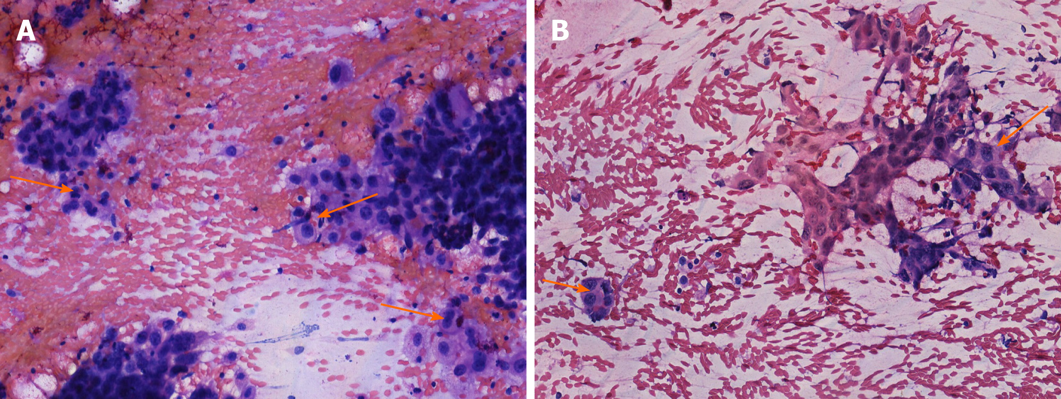
Figure 3 Fine needle aspiration cytology of the thyroid lesions stained with hematoxylin and eosin (× 200).
A: It shows malignant tumor cells (orange arrows); B: It shows malignant tumor cells (orange arrows).
- Citation: Zhang X, Gu X, Li JG, Hu XJ. Metastasis of esophageal squamous cell carcinoma to the thyroid gland with widespread nodal involvement: A case report. World J Clin Cases 2020; 8(19): 4588-4594
- URL: https://www.wjgnet.com/2307-8960/full/v8/i19/4588.htm
- DOI: https://dx.doi.org/10.12998/wjcc.v8.i19.4588


