Copyright
©The Author(s) 2020.
World J Clin Cases. Sep 26, 2020; 8(18): 4272-4279
Published online Sep 26, 2020. doi: 10.12998/wjcc.v8.i18.4272
Published online Sep 26, 2020. doi: 10.12998/wjcc.v8.i18.4272
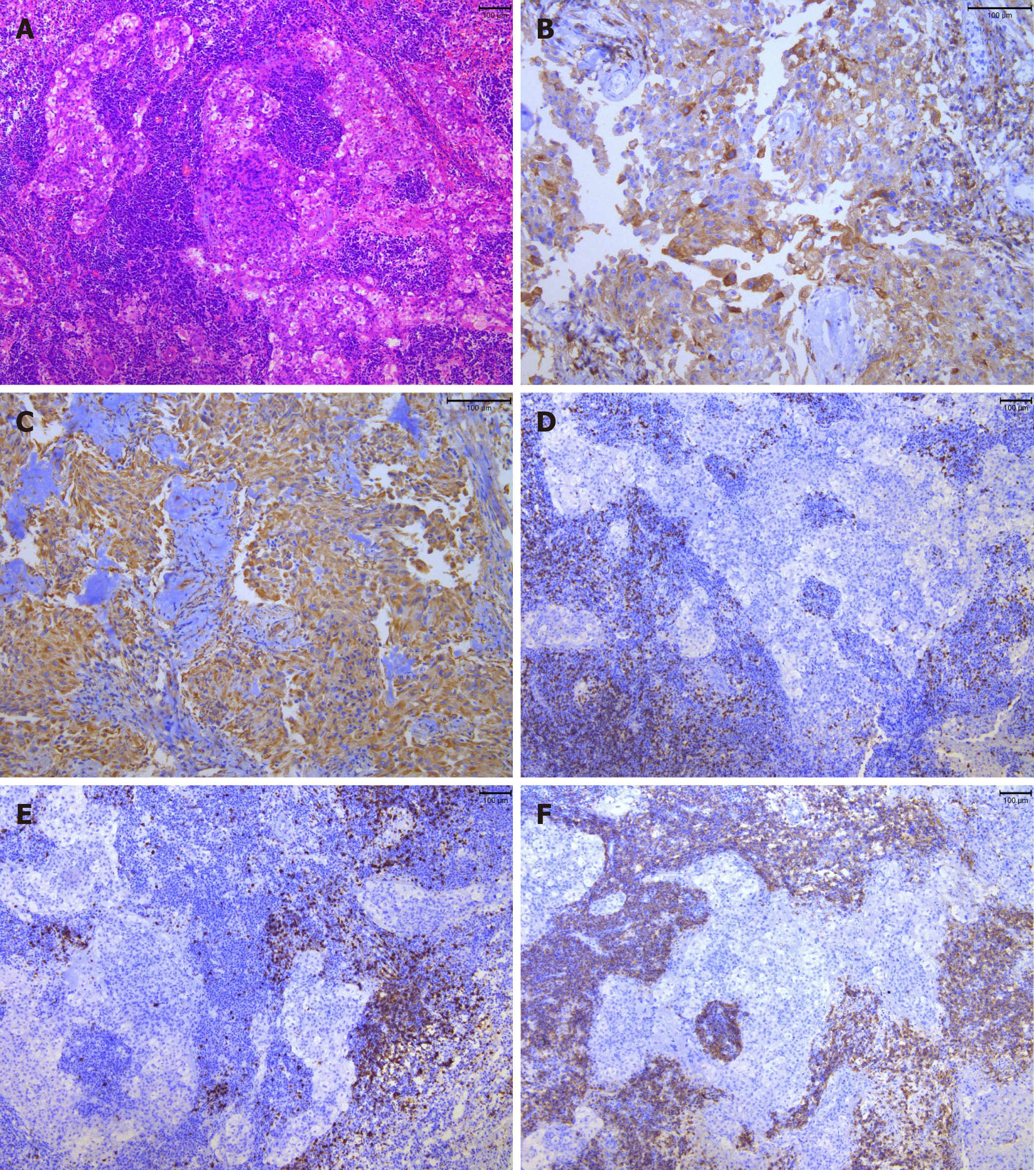
Figure 3 Microscopic characteristics of the tumor tissue.
A: Hematoxylin and eosin staining (100 ×) showed multiple nests of meningothelial cells and syncytial tumor cells arranged in sheets or weaving. The size was consistent, and nuclear heterotypes were not obvious. Tumor stroma with massive lymphocytic and plasmocytic infiltration, and lymphoid follicular formation was identified in a local focus; B: Immunohistochemical staining revealed that the tumor cells were positive for epithelial membrane antigen (200 ×); C: Vimentin (200 ×); D: T lymphocytes were positive for CD3 (100 ×); E: B lymphocytes were positive for CD20 (100 ×); F: Plasmocytes were positive for CD38 (100 ×).
- Citation: Gu KC, Wan Y, Xiang L, Wang LS, Yao WJ. Lymphoplasmacyte-rich meningioma with atypical cystic-solid feature: A case report. World J Clin Cases 2020; 8(18): 4272-4279
- URL: https://www.wjgnet.com/2307-8960/full/v8/i18/4272.htm
- DOI: https://dx.doi.org/10.12998/wjcc.v8.i18.4272


