Copyright
©The Author(s) 2020.
World J Clin Cases. Sep 26, 2020; 8(18): 3978-3987
Published online Sep 26, 2020. doi: 10.12998/wjcc.v8.i18.3978
Published online Sep 26, 2020. doi: 10.12998/wjcc.v8.i18.3978
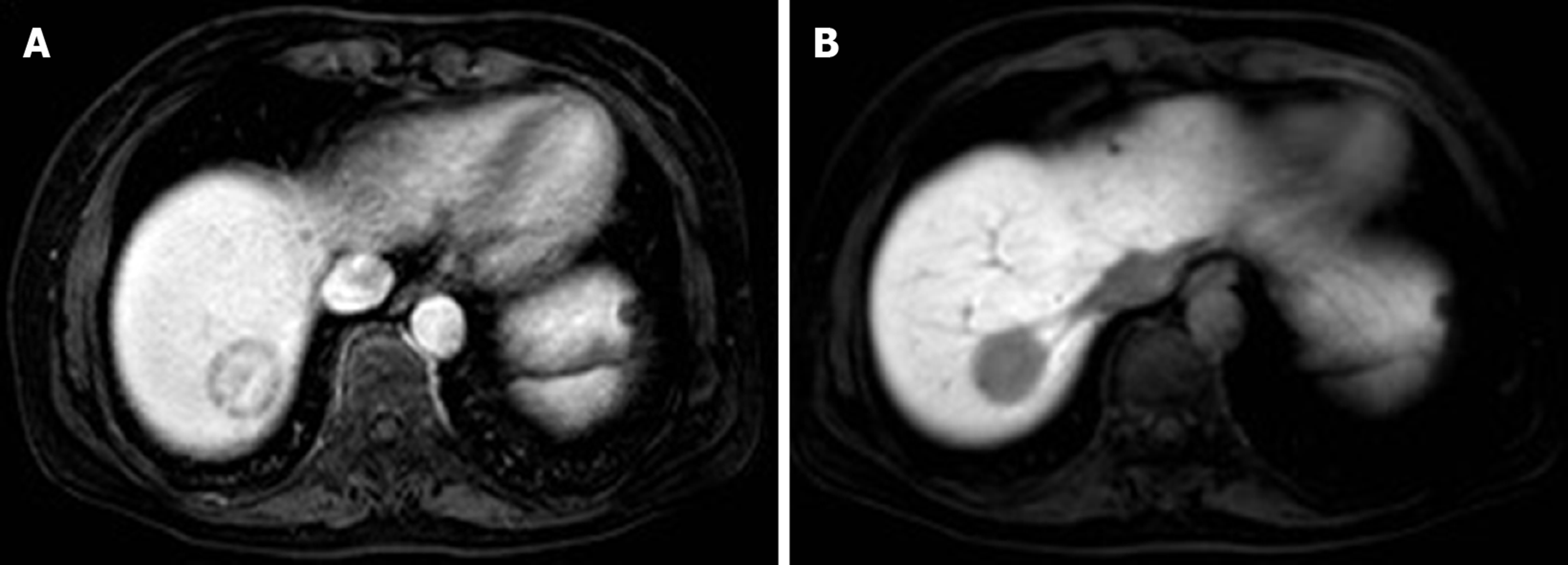
Figure 1 Magnetic resonance imaging.
A: Magnetic resonance imaging shows a target-like sign as a gradual peripheral ring-like enhancement pattern with central low signal intensity in the arterial to delay phases, and the enhanced lesion is surrounded by a thin, hypointense ring on contrasted-enhanced T1-weighted image; B: The lollipop sign consists of a clear tumor mass (candy in the lollipop) and adjacent occluded vein (rod).
- Citation: Kou K, Chen YG, Zhou JP, Sun XD, Sun DW, Li SX, Lv GY. Hepatic epithelioid hemangioendothelioma: Update on diagnosis and therapy. World J Clin Cases 2020; 8(18): 3978-3987
- URL: https://www.wjgnet.com/2307-8960/full/v8/i18/3978.htm
- DOI: https://dx.doi.org/10.12998/wjcc.v8.i18.3978


