Copyright
©The Author(s) 2020.
World J Clin Cases. Sep 6, 2020; 8(17): 3890-3902
Published online Sep 6, 2020. doi: 10.12998/wjcc.v8.i17.3890
Published online Sep 6, 2020. doi: 10.12998/wjcc.v8.i17.3890
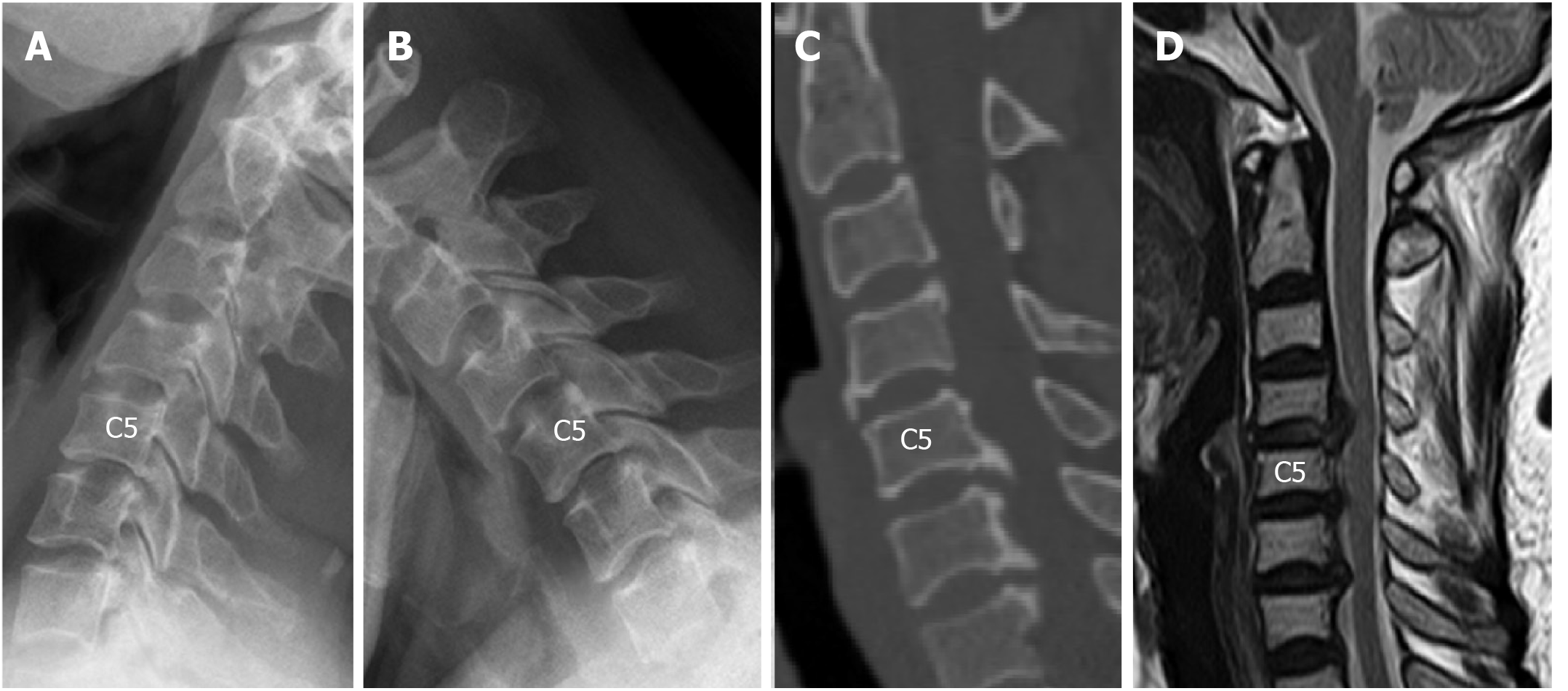
Figure 2 Computed tomography images of case 2.
A and B: Lateral radiographs taken before surgery revealed that the range of motion at C5/6 was decreased (2.57°), while the range of motion at C4/5 and C6/7 was 9.34° and 6.05°, respectively. The intervertebral disc height at C4/5 was preserved; C: Large numbers of osteophytes were revealed at the posterior-inferior border of C5 and C6; D: Magnetic resonance imaging indicated spinal cord compression at C4/5, C5/6, and C6/7, and compression at C4/5 was caused by disc herniation.
- Citation: Wang XF, Meng Y, Liu H, Hong Y, Wang BY. Surgical strategy used in multilevel cervical disc replacement and cervical hybrid surgery: Four case reports. World J Clin Cases 2020; 8(17): 3890-3902
- URL: https://www.wjgnet.com/2307-8960/full/v8/i17/3890.htm
- DOI: https://dx.doi.org/10.12998/wjcc.v8.i17.3890


