Copyright
©The Author(s) 2020.
World J Clin Cases. Sep 6, 2020; 8(17): 3890-3902
Published online Sep 6, 2020. doi: 10.12998/wjcc.v8.i17.3890
Published online Sep 6, 2020. doi: 10.12998/wjcc.v8.i17.3890
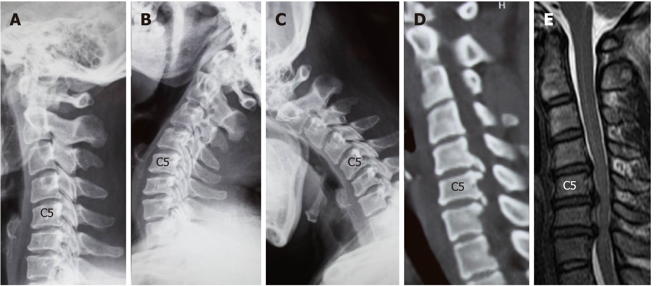
Figure 1 Computed tomography images of case 1.
A-C: Lateral radiographs showing a straightened cervical alignment that returned during flexion-extension movement. The range of motion at C4/5, C5/6, and C6/7 was 11.3°, 14.53°, and 1.28°, respectively; D: Computed tomography image showing large numbers of osteophytes at the posterior border of C5/6 and C6/7; E: Magnetic resonance imaging revealed cervical disc herniation at C4/5 and C6/7.
- Citation: Wang XF, Meng Y, Liu H, Hong Y, Wang BY. Surgical strategy used in multilevel cervical disc replacement and cervical hybrid surgery: Four case reports. World J Clin Cases 2020; 8(17): 3890-3902
- URL: https://www.wjgnet.com/2307-8960/full/v8/i17/3890.htm
- DOI: https://dx.doi.org/10.12998/wjcc.v8.i17.3890


