Copyright
©The Author(s) 2020.
World J Clin Cases. Sep 6, 2020; 8(17): 3821-3827
Published online Sep 6, 2020. doi: 10.12998/wjcc.v8.i17.3821
Published online Sep 6, 2020. doi: 10.12998/wjcc.v8.i17.3821
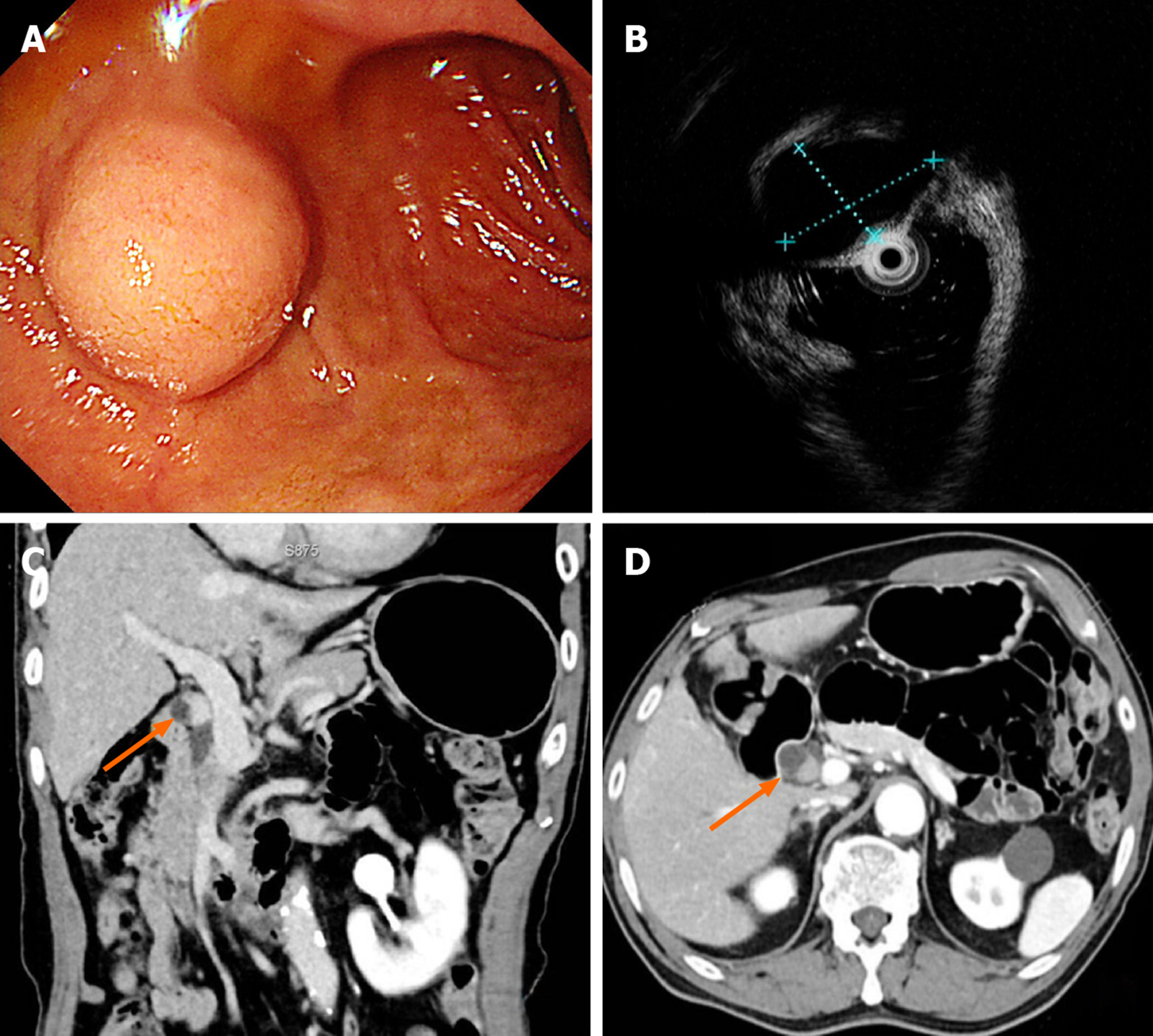
Figure 1 Preoperative tumor evaluation.
A: Upper gastrointestinal endoscopy showing a lesion protruding into the lumen of the duodenal bulb; B: Endoscopic ultrasonography showing a hypoechoic lesion 1.8 cm in size; C: Coronary view of abdominal computed tomography (CT) showing a small enhancing nodule 1.4 cm in size (orange arrow) between the cystic duct and the duodenal bulb; D: Axial view of abdominal CT showing the lesion (orange arrow).
- Citation: Kim DH, Park JH, Cho JK, Yang JW, Kim TH, Jeong SH, Kim YH, Lee YJ, Hong SC, Jung EJ, Ju YT, Jeong CY, Kim JY. Traumatic neuroma of remnant cystic duct mimicking duodenal subepithelial tumor: A case report. World J Clin Cases 2020; 8(17): 3821-3827
- URL: https://www.wjgnet.com/2307-8960/full/v8/i17/3821.htm
- DOI: https://dx.doi.org/10.12998/wjcc.v8.i17.3821


