Copyright
©The Author(s) 2020.
World J Clin Cases. Sep 6, 2020; 8(17): 3751-3762
Published online Sep 6, 2020. doi: 10.12998/wjcc.v8.i17.3751
Published online Sep 6, 2020. doi: 10.12998/wjcc.v8.i17.3751
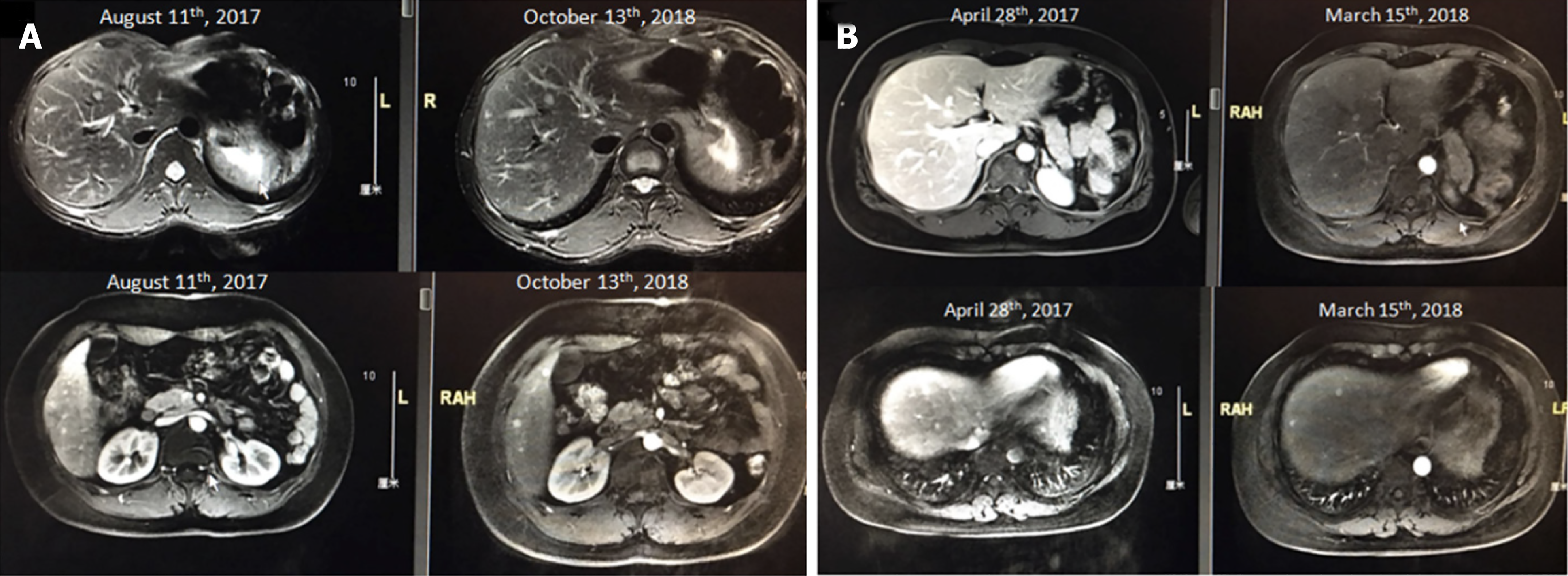
Figure 2 Magnetic resonance imaging screening showing an example of the largest axis of a liver metastasis < 5 mm.
A: Patient MRI scan image in 2017 August and 2018 October. B: Patient magnetic resonance imaging (MRI) scan image in 2017 April and 2018 March.
- Citation: Gao HL, Wang WQ, Xu HX, Wu CT, Li H, Ni QX, Yu XJ, Liu L. Active surveillance in metastatic pancreatic neuroendocrine tumors: A 20-year single-institutional experience. World J Clin Cases 2020; 8(17): 3751-3762
- URL: https://www.wjgnet.com/2307-8960/full/v8/i17/3751.htm
- DOI: https://dx.doi.org/10.12998/wjcc.v8.i17.3751


