Copyright
©The Author(s) 2020.
World J Clin Cases. Aug 26, 2020; 8(16): 3591-3600
Published online Aug 26, 2020. doi: 10.12998/wjcc.v8.i16.3591
Published online Aug 26, 2020. doi: 10.12998/wjcc.v8.i16.3591
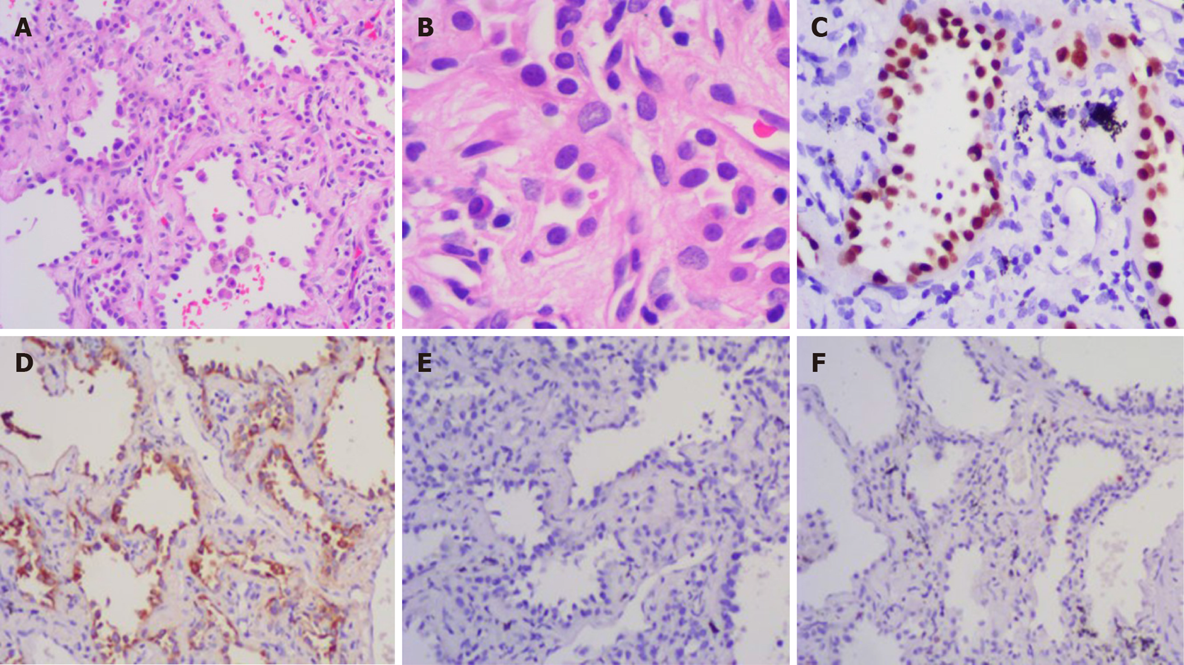
Figure 5 The tissue section showing lung adenocarcinoma.
A: (100 ×) Pathological examination indicated lung adenocarcinoma; B: (400 ×) Tumour cells showed prominent atypia at high magnification; C: (200 ×) Thyroid transcription factor-1 was positive; D: (200 ×) Napsin-A was positive; E: GCDFP-15 was negative; and F: Ki-67 was focally positive.
- Citation: Zhang T, Feng L, Lian J, Ren WL. Giant benign phyllodes breast tumour with pulmonary nodule mimicking malignancy: A case report. World J Clin Cases 2020; 8(16): 3591-3600
- URL: https://www.wjgnet.com/2307-8960/full/v8/i16/3591.htm
- DOI: https://dx.doi.org/10.12998/wjcc.v8.i16.3591


