Copyright
©The Author(s) 2020.
World J Clin Cases. Aug 26, 2020; 8(16): 3591-3600
Published online Aug 26, 2020. doi: 10.12998/wjcc.v8.i16.3591
Published online Aug 26, 2020. doi: 10.12998/wjcc.v8.i16.3591
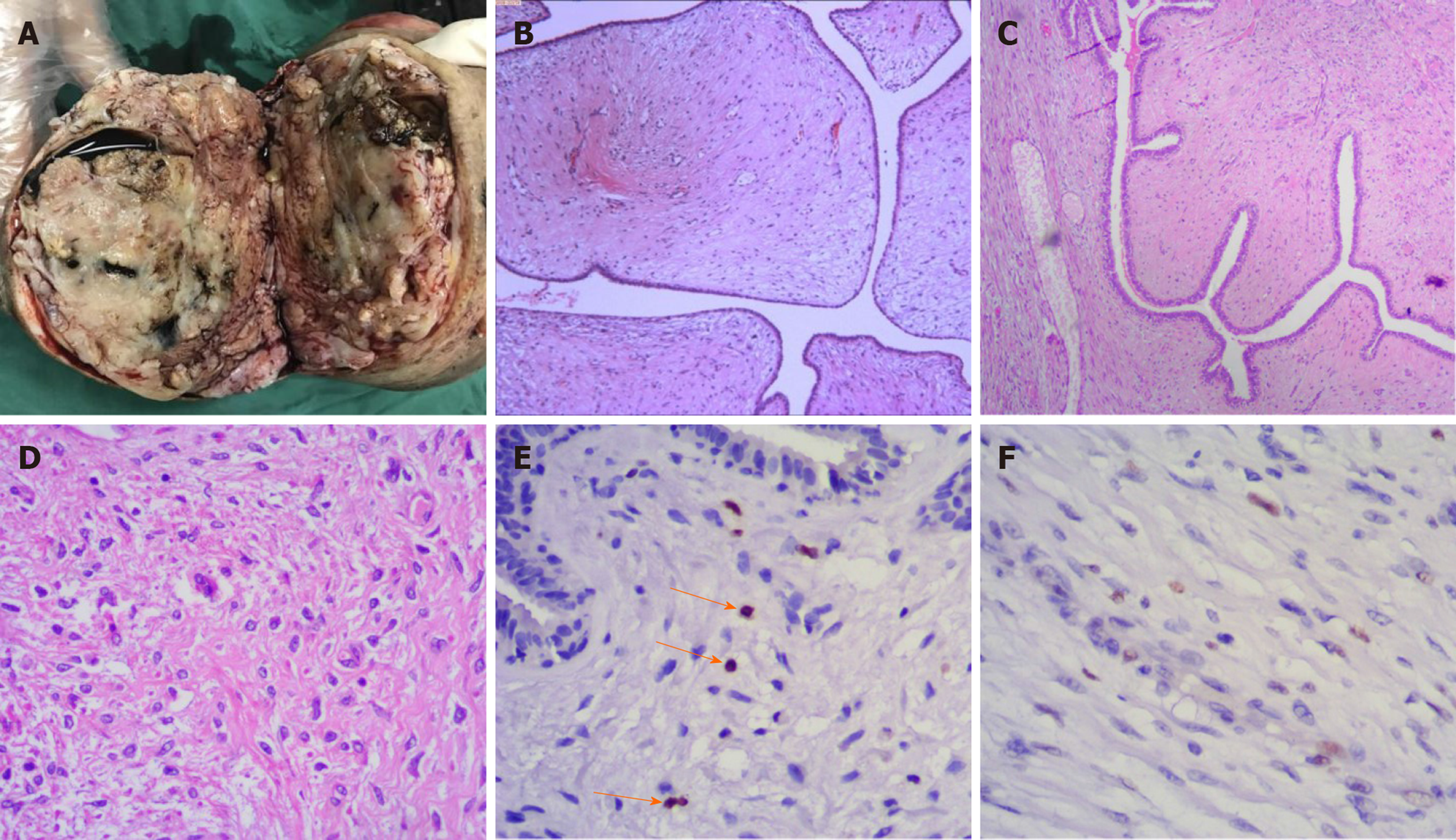
Figure 4 The tissue section showing benign phyllodes tumor.
A: Cystic components after incision of the tumour; B: (10 ×) Well-circumscribed fibroepithelial neoplasm; C: (40 ×) Prominent leaf-like architecture and areas of hypocellular stroma; D: (400 ×) Bland stromal spindle cells without mitoses or nuclear atypia; E: Ki-67 proliferation index of the tumour was 1 for the stromal component; and F: The P53 index of the stromal component was focally positive.
- Citation: Zhang T, Feng L, Lian J, Ren WL. Giant benign phyllodes breast tumour with pulmonary nodule mimicking malignancy: A case report. World J Clin Cases 2020; 8(16): 3591-3600
- URL: https://www.wjgnet.com/2307-8960/full/v8/i16/3591.htm
- DOI: https://dx.doi.org/10.12998/wjcc.v8.i16.3591


