Copyright
©The Author(s) 2020.
World J Clin Cases. Aug 26, 2020; 8(16): 3583-3590
Published online Aug 26, 2020. doi: 10.12998/wjcc.v8.i16.3583
Published online Aug 26, 2020. doi: 10.12998/wjcc.v8.i16.3583
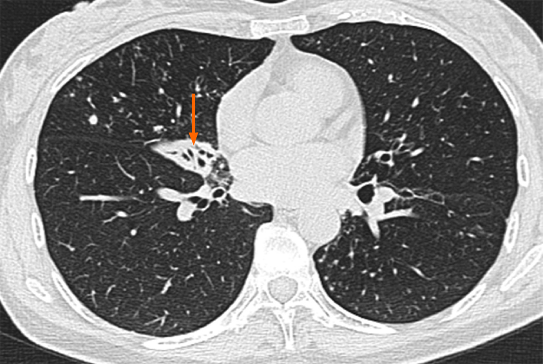
Figure 2 Another computed tomography image obtained in March 2018 revealed that the bronchiectasis in the middle lobe of her right lung was accompanied by atelectasis (arrow), which was more observable than the atrophy in the same location in the previous computed tomography image.
- Citation: Han XY, Wang YY, Wei HQ, Yang GZ, Wang J, Jia YZ, Ao WQ. Multifocal neuroendocrine cell hyperplasia accompanied by tumorlet formation and pulmonary sclerosing pneumocytoma: A case report. World J Clin Cases 2020; 8(16): 3583-3590
- URL: https://www.wjgnet.com/2307-8960/full/v8/i16/3583.htm
- DOI: https://dx.doi.org/10.12998/wjcc.v8.i16.3583


