Copyright
©The Author(s) 2020.
World J Clin Cases. Aug 26, 2020; 8(16): 3583-3590
Published online Aug 26, 2020. doi: 10.12998/wjcc.v8.i16.3583
Published online Aug 26, 2020. doi: 10.12998/wjcc.v8.i16.3583
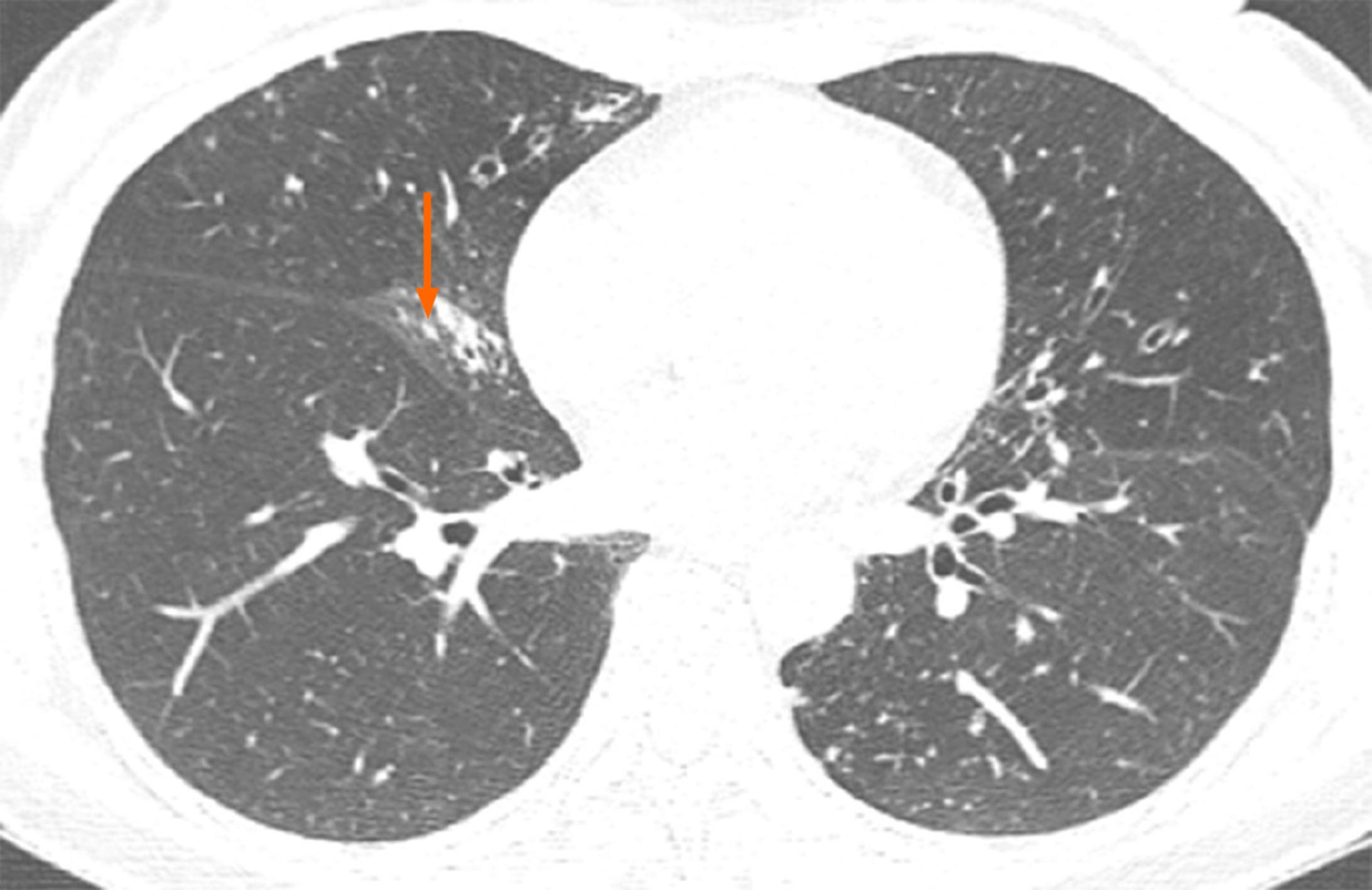
Figure 1 In November 2017, computed tomography showed atrophy in the middle lobe of her right lung as well as bronchiectasis in the left lung and the middle lobe of the right lung accompanied by infections.
Numerous nodules with a diameter of 0.3 to 0.5 cm were identified in the middle lobe of the right lung (arrow).
- Citation: Han XY, Wang YY, Wei HQ, Yang GZ, Wang J, Jia YZ, Ao WQ. Multifocal neuroendocrine cell hyperplasia accompanied by tumorlet formation and pulmonary sclerosing pneumocytoma: A case report. World J Clin Cases 2020; 8(16): 3583-3590
- URL: https://www.wjgnet.com/2307-8960/full/v8/i16/3583.htm
- DOI: https://dx.doi.org/10.12998/wjcc.v8.i16.3583


