Copyright
©The Author(s) 2020.
World J Clin Cases. Aug 26, 2020; 8(16): 3548-3552
Published online Aug 26, 2020. doi: 10.12998/wjcc.v8.i16.3548
Published online Aug 26, 2020. doi: 10.12998/wjcc.v8.i16.3548
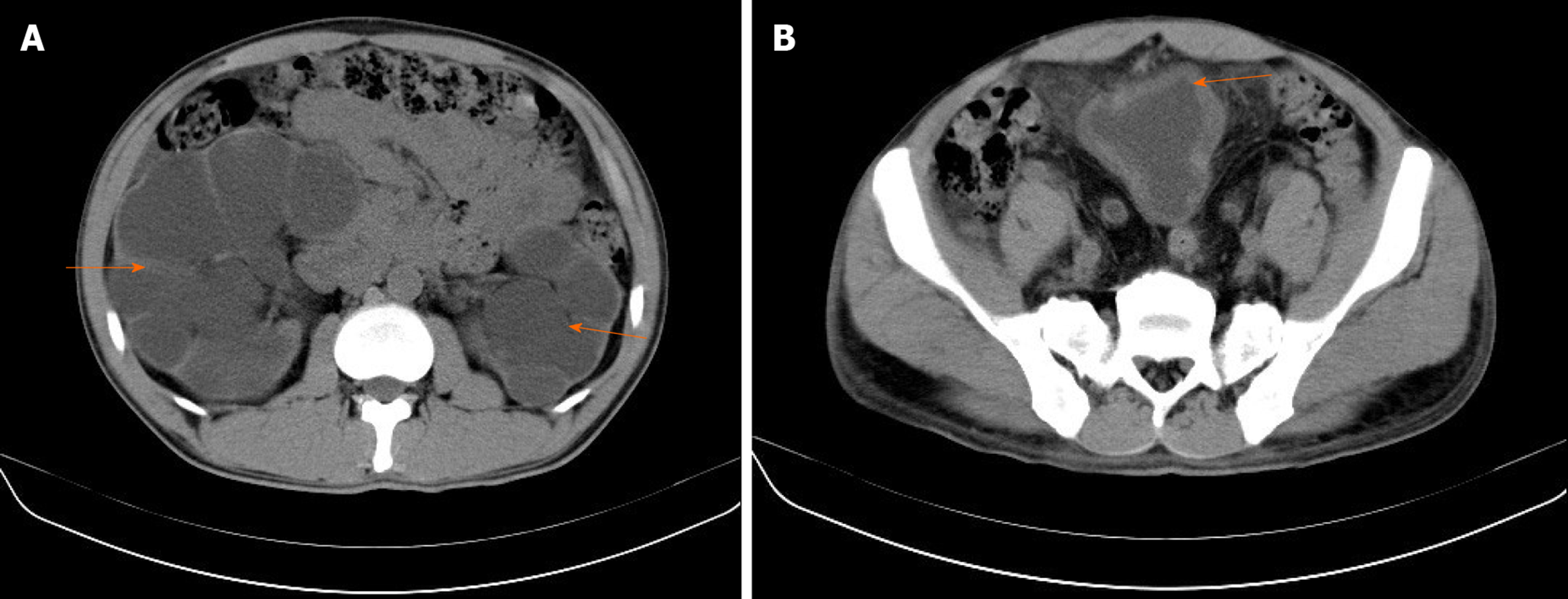
Figure 1 Computed tomography examination.
A: Computed tomography showed bilateral hydronephrosis (orange arrow); B: A thick walled urinary bladder (orange arrow) with increased fat density around the bladder.
- Citation: Zhao J, Fu YX, Feng G, Mo CB. Pelvic lipomatosis and renal transplantation: A case report. World J Clin Cases 2020; 8(16): 3548-3552
- URL: https://www.wjgnet.com/2307-8960/full/v8/i16/3548.htm
- DOI: https://dx.doi.org/10.12998/wjcc.v8.i16.3548


