
















©The Author(s) 2020.
World J Clin Cases. Aug 6, 2020; 8(15): 3341-3348
Published online Aug 6, 2020. doi: 10.12998/wjcc.v8.i15.3341
Published online Aug 6, 2020. doi: 10.12998/wjcc.v8.i15.3341
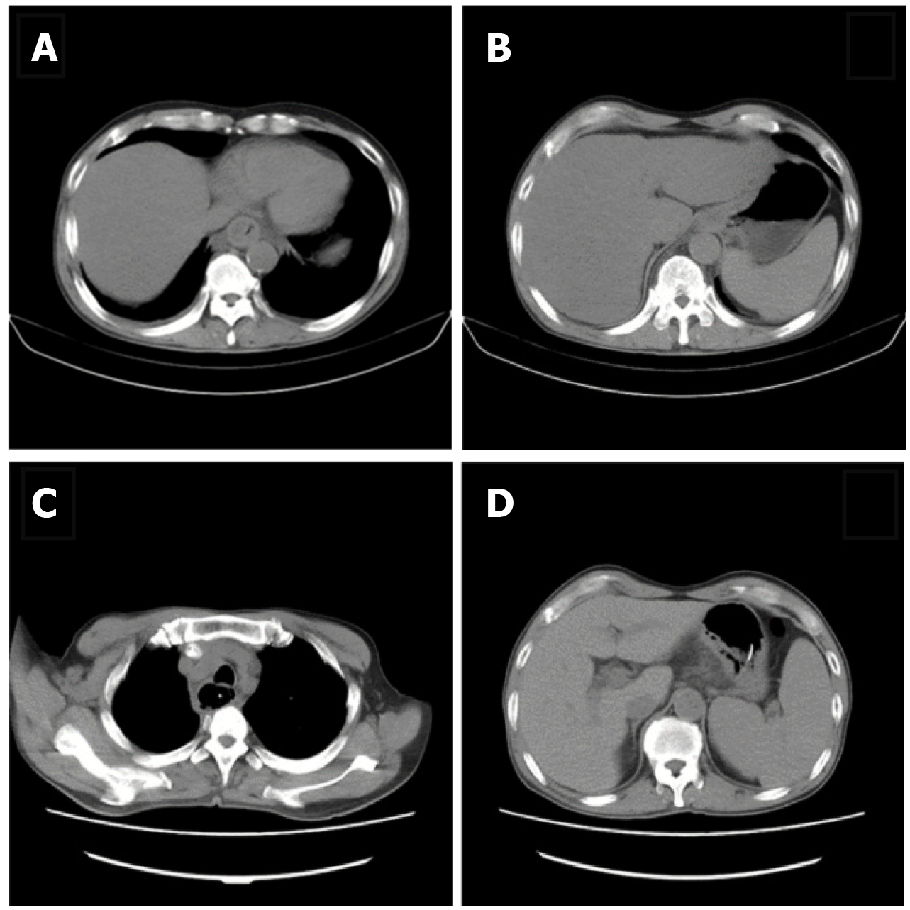
Figure 1 Thoracic computed tomography.
A: Thoracic computed tomography (CT) scan showed oesophageal dilatation and effusion, wall thickening of the lower oesophagus, inflammation of the upper lobe of the right lung, and bilateral pleural effusion; B: Abdominal CT scan showed thickening of the gastric wall of the cardia and adjacent lesser curvature of the stomach, fatty liver, and gallstones; C: Chest CT scan re-examination revealed oesophageal mucosal exfoliation involving the stomach wall. Right upper and middle lobe inflammation had improved since the previous report and oesophageal dilatation and effusion had reduced; D: Abdominal CT scan showed thickening of the gastric wall of the cardia and adjacent lesser curvature of the stomach which was the same as the previous review.
- Citation: Men CJ, Singh SK, Zhang GL, Wang Y, Liu CW. Acute suppurative oesophagitis with fever and cough: A case report. World J Clin Cases 2020; 8(15): 3341-3348
- URL: https://www.wjgnet.com/2307-8960/full/v8/i15/3341.htm
- DOI: https://dx.doi.org/10.12998/wjcc.v8.i15.3341