
















©The Author(s) 2020.
World J Clin Cases. Aug 6, 2020; 8(15): 3177-3187
Published online Aug 6, 2020. doi: 10.12998/wjcc.v8.i15.3177
Published online Aug 6, 2020. doi: 10.12998/wjcc.v8.i15.3177
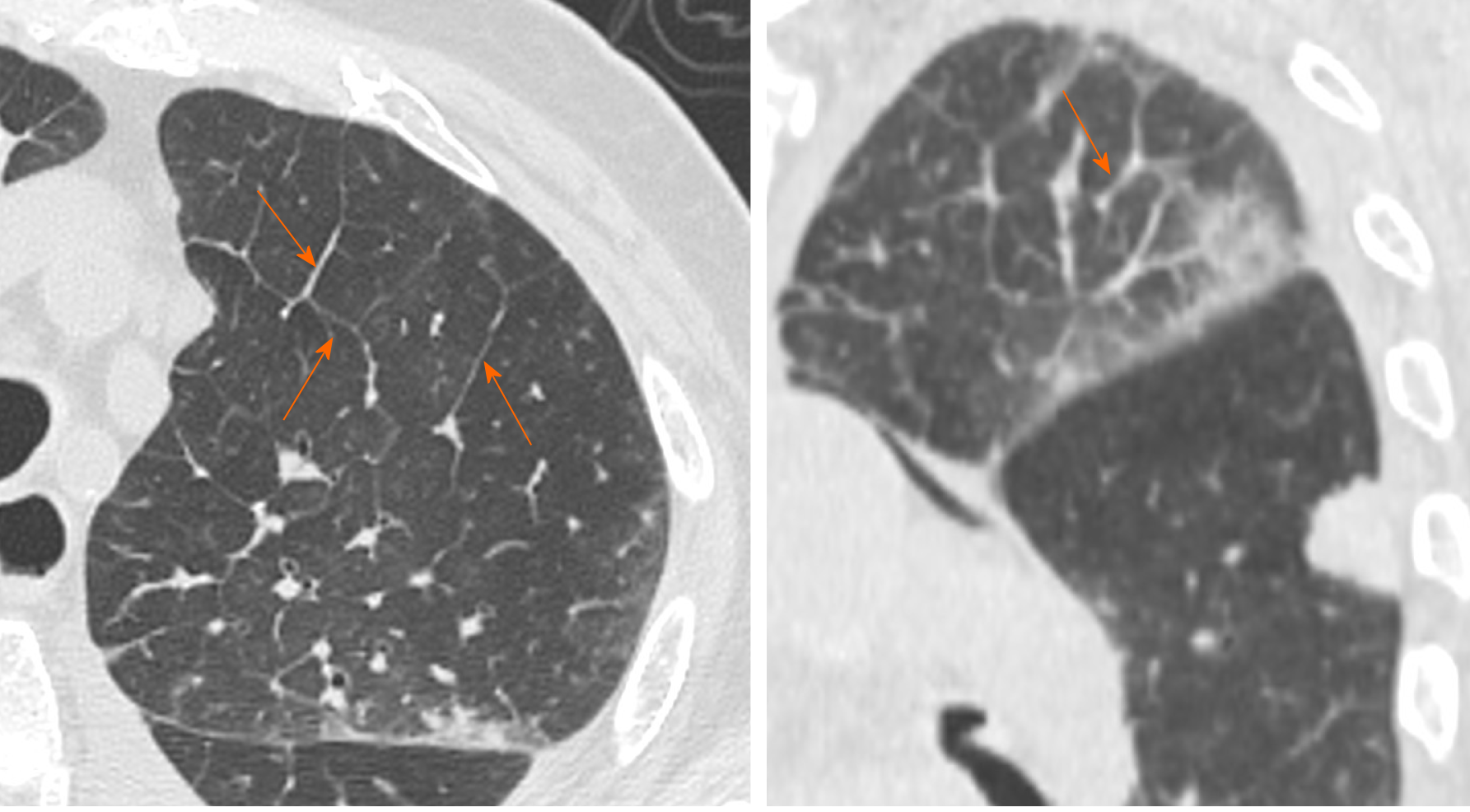
Figure 7 Computed tomography images of interstitial septal thickening.
92-year man admitted to the emergency room presenting cough, fever and worsening dyspnea. Chest computed tomography showed interlobular septal thickening predominantly in left upper lobe (orange arrows).
- Citation: Caruso D, Polidori T, Guido G, Nicolai M, Bracci B, Cremona A, Zerunian M, Polici M, Pucciarelli F, Rucci C, Dominicis CD, Girolamo MD, Argento G, Sergi D, Laghi A. Typical and atypical COVID-19 computed tomography findings. World J Clin Cases 2020; 8(15): 3177-3187
- URL: https://www.wjgnet.com/2307-8960/full/v8/i15/3177.htm
- DOI: https://dx.doi.org/10.12998/wjcc.v8.i15.3177