Copyright
©The Author(s) 2020.
World J Clin Cases. Jun 26, 2020; 8(12): 2647-2654
Published online Jun 26, 2020. doi: 10.12998/wjcc.v8.i12.2647
Published online Jun 26, 2020. doi: 10.12998/wjcc.v8.i12.2647
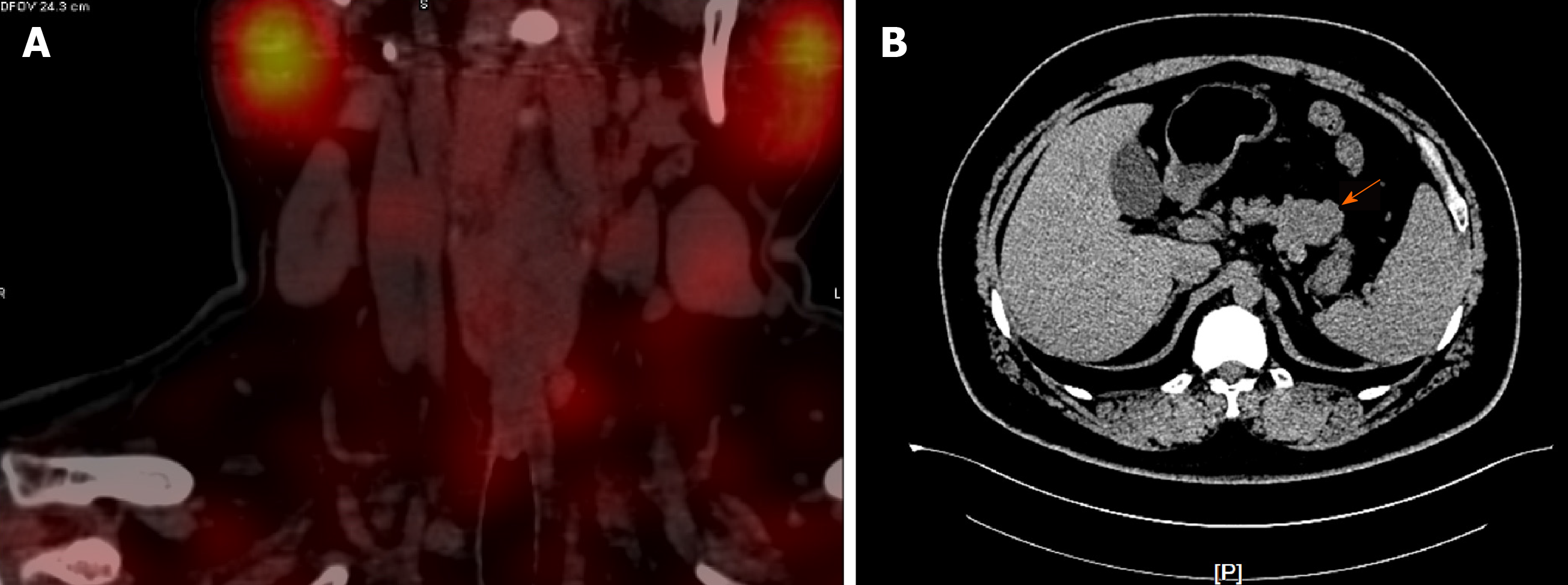
Figure 1 Computed tomography examination.
A: Parathyroid two-phase computed tomography revealed a strong signal in the upper part of the left lobes and posterior part of the right lobes of the thyroid; B: Pancreatic perfusion computed tomography imaging revealed irregular soft tissue densities of the pancreas body, which were localized around the stomach wall.
- Citation: Ma CH, Guo HB, Pan XY, Zhang WX. Comprehensive treatment of rare multiple endocrine neoplasia type 1: A case report. World J Clin Cases 2020; 8(12): 2647-2654
- URL: https://www.wjgnet.com/2307-8960/full/v8/i12/2647.htm
- DOI: https://dx.doi.org/10.12998/wjcc.v8.i12.2647


