Copyright
©The Author(s) 2020.
World J Clin Cases. Jun 6, 2020; 8(11): 2350-2358
Published online Jun 6, 2020. doi: 10.12998/wjcc.v8.i11.2350
Published online Jun 6, 2020. doi: 10.12998/wjcc.v8.i11.2350
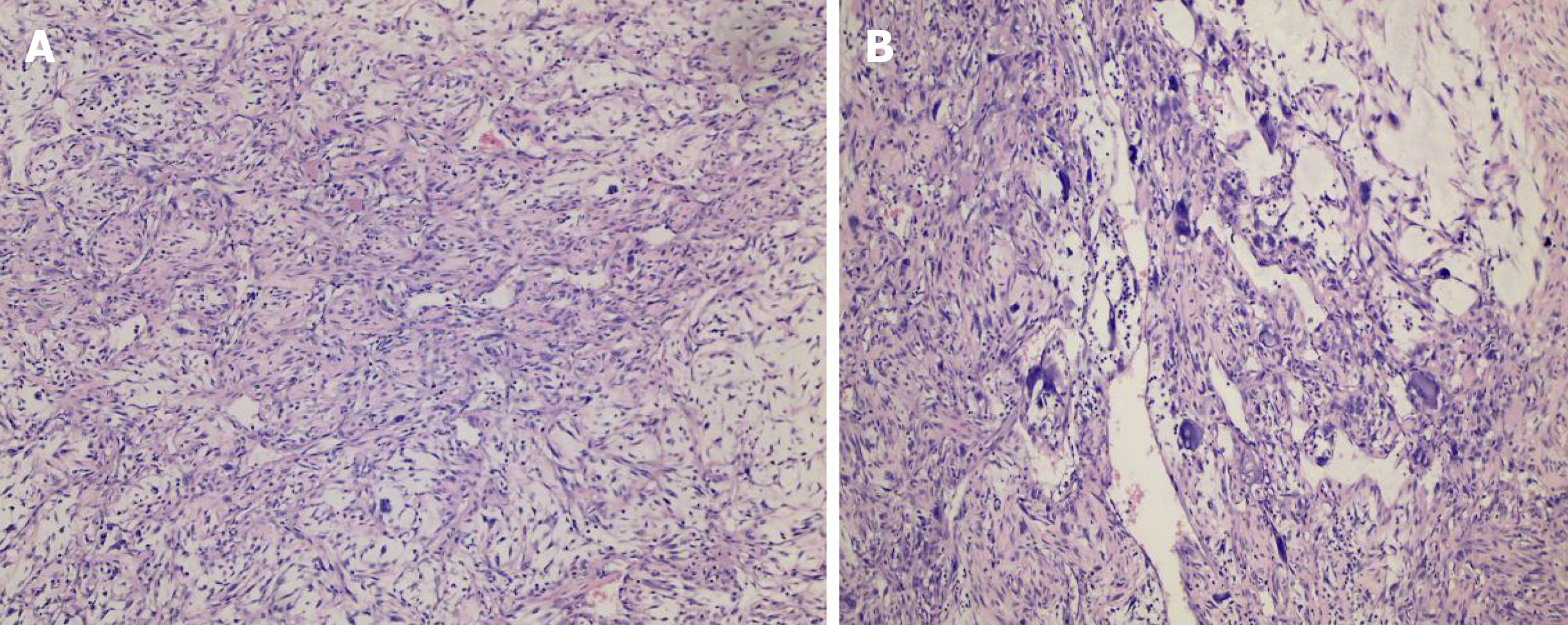
Figure 4 Pathologic images of the mass.
Histologic evaluation showed that there were abundant heteromorphic spindle cells and partial nodular mucus, arranged in a woven pattern, with rare nuclear fission and abundant blood vessels in the interstitium (A: Hematoxylin-eosin staining, × 100, B: Hematoxylin-eosin staining, × 200).
- Citation: Ke XT, Yu XF, Liu JY, Huang F, Chen MG, Lai QQ. Myxofibrosarcoma of the scalp with difficult preoperative diagnosis: A case report and review of the literature. World J Clin Cases 2020; 8(11): 2350-2358
- URL: https://www.wjgnet.com/2307-8960/full/v8/i11/2350.htm
- DOI: https://dx.doi.org/10.12998/wjcc.v8.i11.2350


