Copyright
©The Author(s) 2020.
World J Clin Cases. Jun 6, 2020; 8(11): 2332-2338
Published online Jun 6, 2020. doi: 10.12998/wjcc.v8.i11.2332
Published online Jun 6, 2020. doi: 10.12998/wjcc.v8.i11.2332
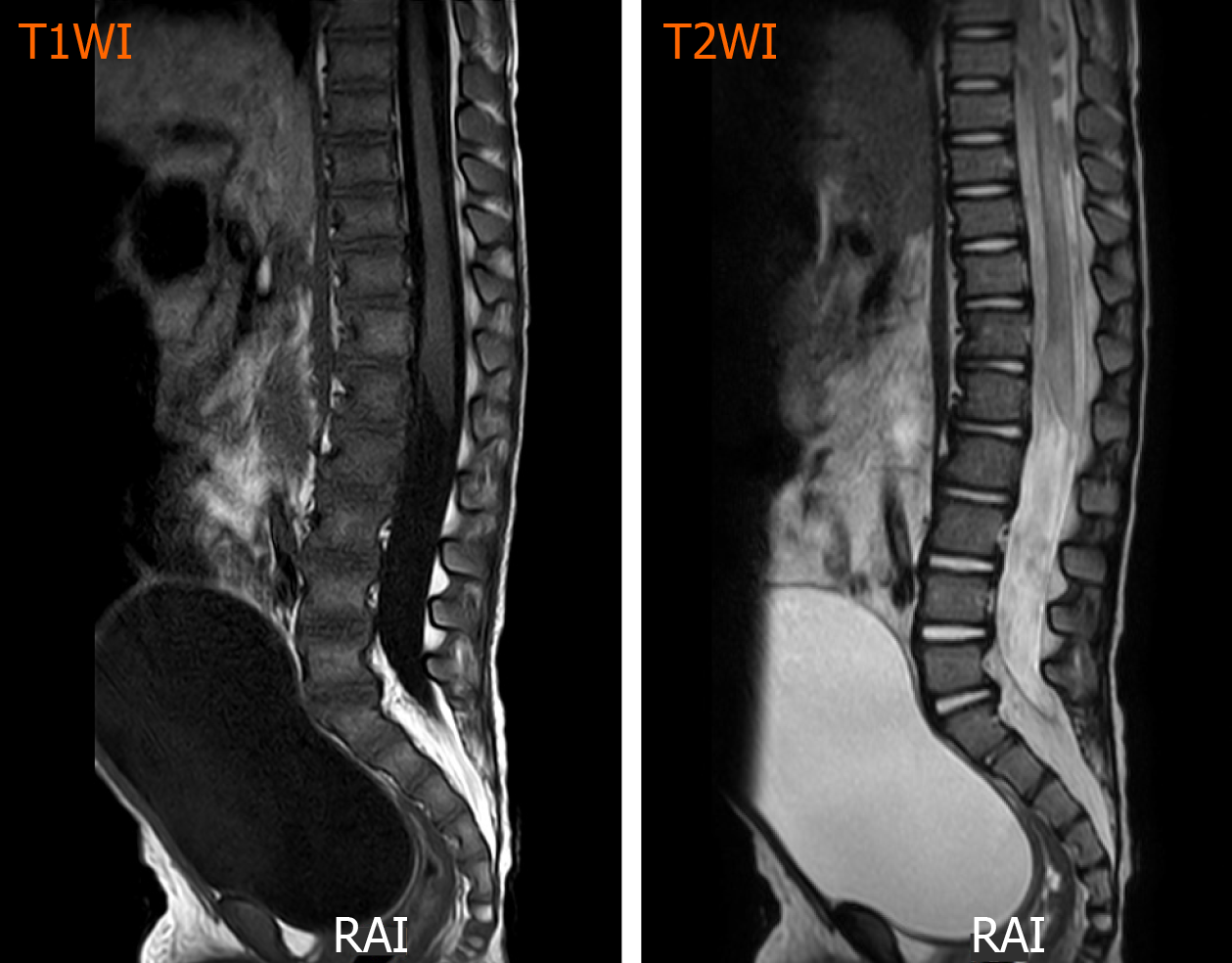
Figure 3 T1/T2-weighted sagittal images of the lumbar spines showing some increased intramedullary signal intensity around the L1-L5 level.
T1WI: T1-weighted sagittal image; T2WI: T2-weighted sagittal image.
- Citation: Cai JB, He M, Wang FL, Xiong JN, Mao JQ, Guan ZH, Li LJ, Wang JH. Paraplegia after transcatheter artery chemoembolization in a child with clear cell sarcoma of the kidney: A case report. World J Clin Cases 2020; 8(11): 2332-2338
- URL: https://www.wjgnet.com/2307-8960/full/v8/i11/2332.htm
- DOI: https://dx.doi.org/10.12998/wjcc.v8.i11.2332


