Copyright
©The Author(s) 2020.
World J Clin Cases. Jun 6, 2020; 8(11): 2325-2331
Published online Jun 6, 2020. doi: 10.12998/wjcc.v8.i11.2325
Published online Jun 6, 2020. doi: 10.12998/wjcc.v8.i11.2325
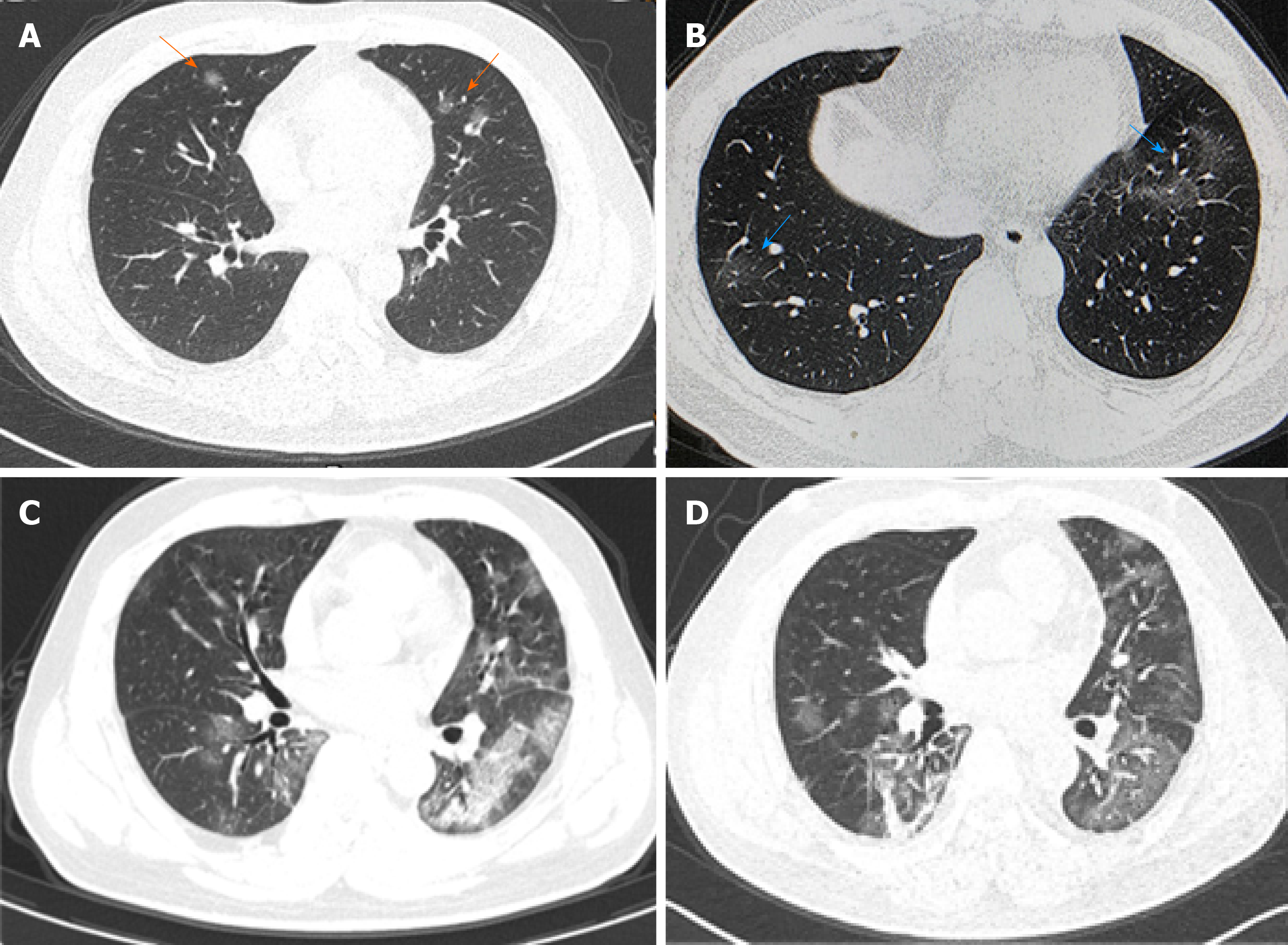
Figure 1 Chest computed tomography results of the coronavirus disease 2019 patient.
A: Computed tomography (CT) results of the chest on January 22 showed a small amount of ground-glass exudation in both lungs (arrows); B: CT results of the chest on January 26 showed an increased amount of exudation in both lungs (arrows); C: CT results of the chest on January 31 showed a large amount of exudation in both lungs which were more prominent in the left lung; D: CT results of the chest on February 3 showed that bilateral lung exudation was absorbed and local fibrosis was identified.
- Citation: He YF, Lian SJ, Dong YC. Clinical characteristics, diagnosis, and treatment of COVID-19: A case report. World J Clin Cases 2020; 8(11): 2325-2331
- URL: https://www.wjgnet.com/2307-8960/full/v8/i11/2325.htm
- DOI: https://dx.doi.org/10.12998/wjcc.v8.i11.2325


