Copyright
©The Author(s) 2020 Published by Baishideng Publishing Group Inc.
World J Clin Cases. Jun 6, 2020; 8(11): 2294-2304
Published online Jun 6, 2020. doi: 10.12998/wjcc.v8.i11.2294
Published online Jun 6, 2020. doi: 10.12998/wjcc.v8.i11.2294
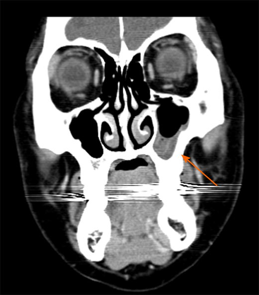
Figure 2 Cone beam computed tomography of patient with maxillary sinusitis (Case 3).
Coronal view showing left maxillary sinus with high density fluid (arrow).
- Citation: Park S, Park JW. Various diagnostic possibilities for zygomatic arch pain: Seven case reports and review of literature. World J Clin Cases 2020; 8(11): 2294-2304
- URL: https://www.wjgnet.com/2307-8960/full/v8/i11/2294.htm
- DOI: https://dx.doi.org/10.12998/wjcc.v8.i11.2294


