
















©The Author(s) 2020.
World J Clin Cases. May 26, 2020; 8(10): 2050-2055
Published online May 26, 2020. doi: 10.12998/wjcc.v8.i10.2050
Published online May 26, 2020. doi: 10.12998/wjcc.v8.i10.2050
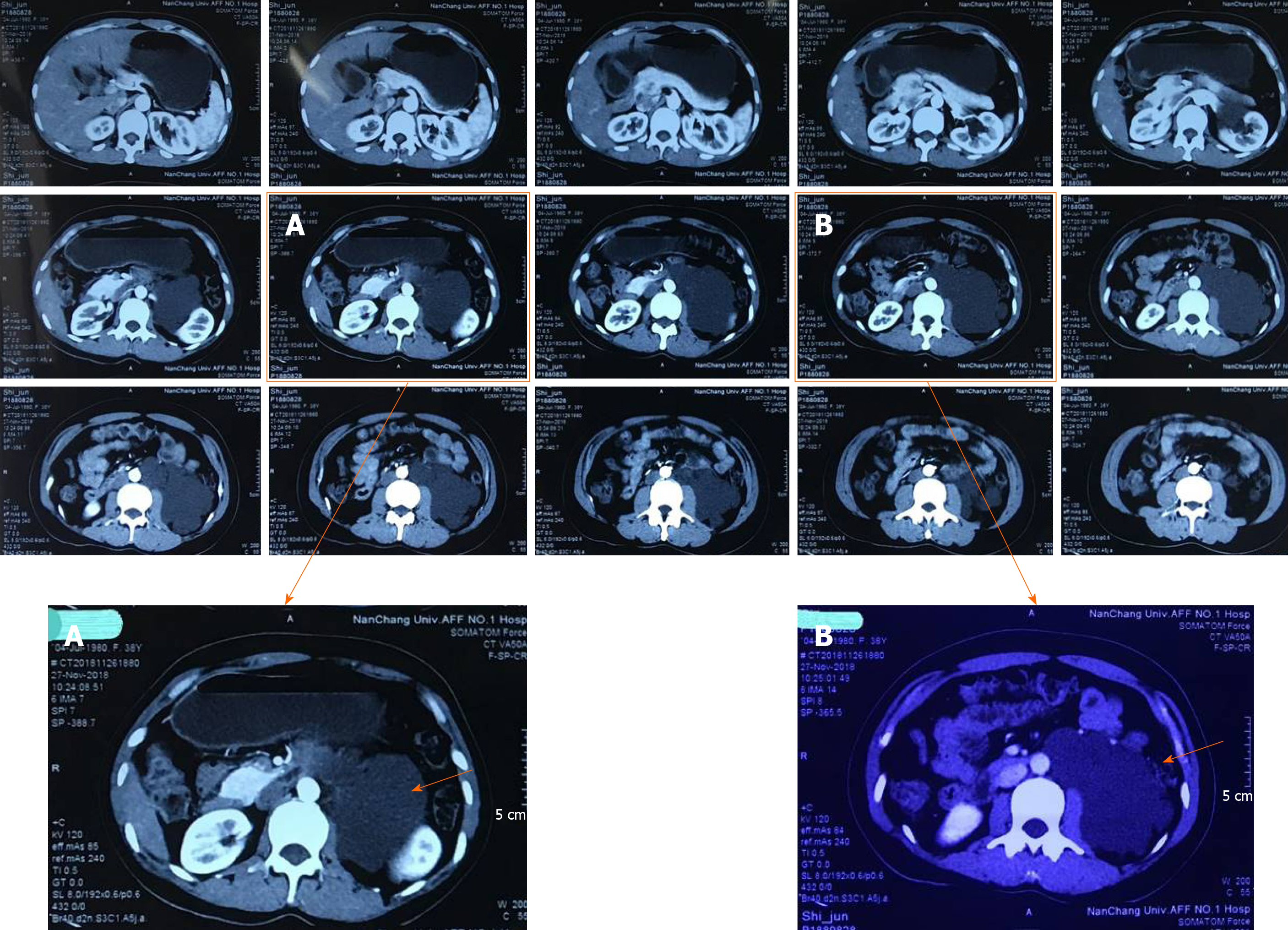
Figure 1 Preoperative angiography of the double renal artery.
A: Cystic low-density shadow in the left renal hilus with a lacy edge; B: Mass adjacent to the abdominal aorta, wrapping the left side, compressing the surrounding tissue, the upper segment of the left ureter, and dilated hydronephrosis in the left kidney.
- Citation: Zhang C, Fu B, Xu S, Zhou XC, Cheng XF, Fu WQ, Wang GX. Robot-assisted retroperitoneal laparoscopic excision of perirenal vascular tumor: A case report. World J Clin Cases 2020; 8(10): 2050-2055
- URL: https://www.wjgnet.com/2307-8960/full/v8/i10/2050.htm
- DOI: https://dx.doi.org/10.12998/wjcc.v8.i10.2050