















©The Author(s) 2020.
World J Clin Cases. May 26, 2020; 8(10): 2016-2022
Published online May 26, 2020. doi: 10.12998/wjcc.v8.i10.2016
Published online May 26, 2020. doi: 10.12998/wjcc.v8.i10.2016
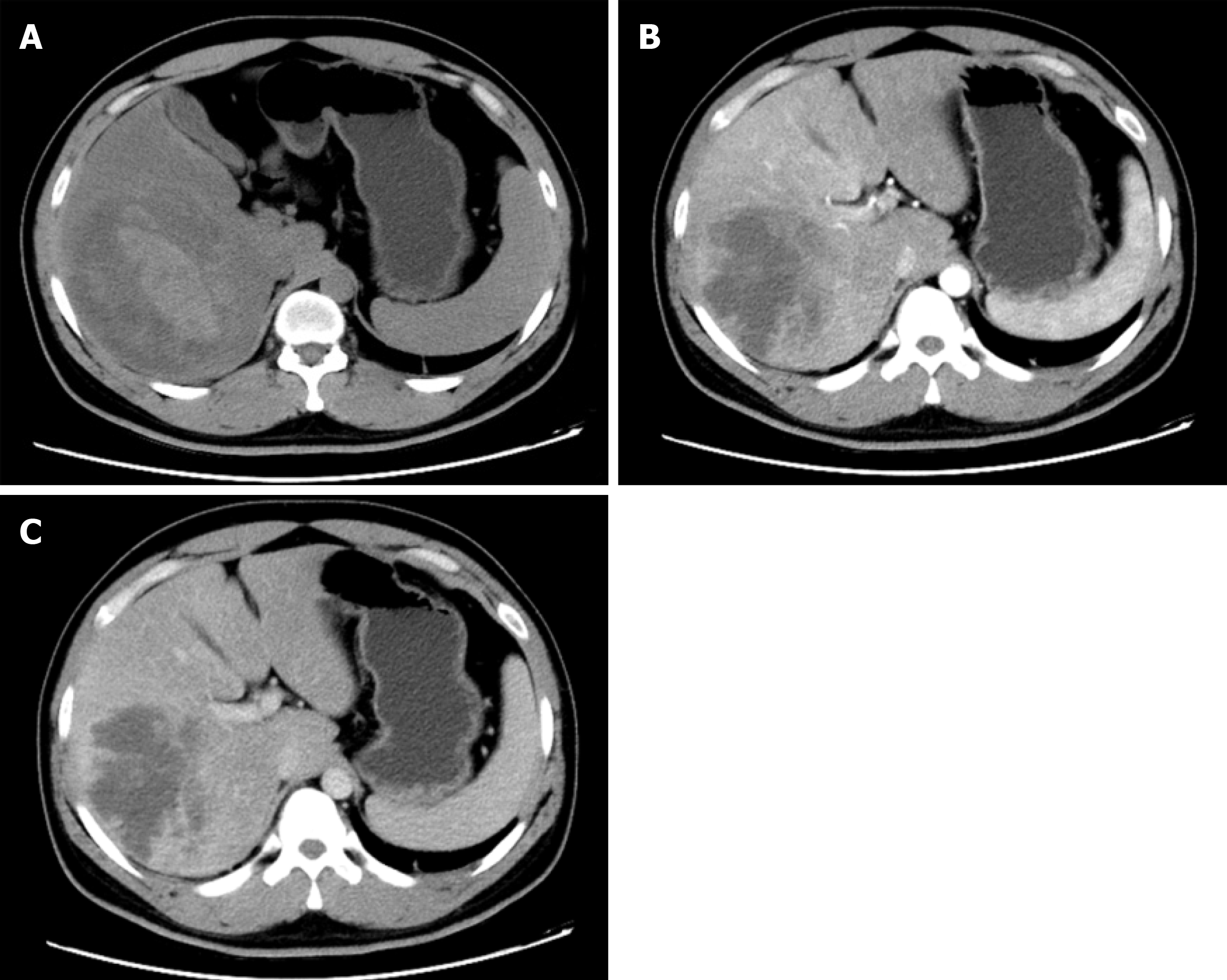
Figure 1 Computed tomography imaging of liver infarction.
A: Plain scan phase of computed tomography (CT) detected an irregular lesion in the right lobe of the liver; B: Arterial phase of CT showed no obvious enhancement; C: Venous phase of CT showed no obvious enhancement.
- Citation: Wang FH, Yang NN, Liu F, Tian H. Unexplained huge liver infarction presenting as a tumor with bleeding: A case report. World J Clin Cases 2020; 8(10): 2016-2022
- URL: https://www.wjgnet.com/2307-8960/full/v8/i10/2016.htm
- DOI: https://dx.doi.org/10.12998/wjcc.v8.i10.2016