Copyright
©The Author(s) 2020.
World J Clin Cases. May 26, 2020; 8(10): 2009-2015
Published online May 26, 2020. doi: 10.12998/wjcc.v8.i10.2009
Published online May 26, 2020. doi: 10.12998/wjcc.v8.i10.2009
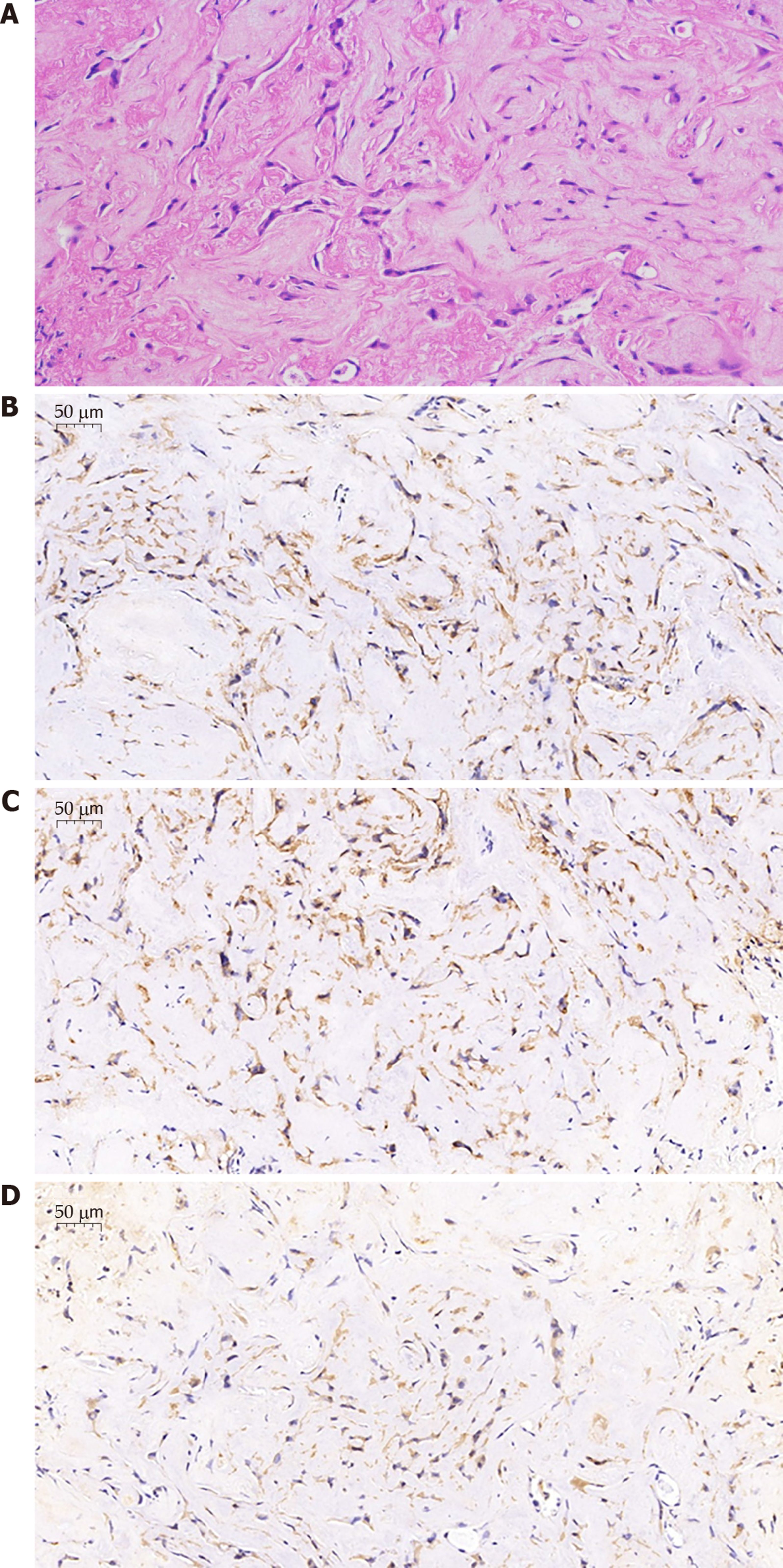
Figure 3 Immunohistochemical findings.
A: Tumor cells, in a short strip form and with epithelial cell features, have no nuclear division and contain abundant eosinophils in the cytoplasm (× 20). B and C: Immunohistochemical analyses for CD34 (B) and CD31 (C) are positive both in the cytoplasm and the tumor cell membrane (× 40); D: Immunohistochemical analysis for Vimentin reveals positivity in the cytoplasm (× 40).
- Citation: Zhang XQ, Chen H, Song S, Qin Y, Cai LM, Zhang F. Effective combined therapy for pulmonary epithelioid hemangioendothelioma: A case report. World J Clin Cases 2020; 8(10): 2009-2015
- URL: https://www.wjgnet.com/2307-8960/full/v8/i10/2009.htm
- DOI: https://dx.doi.org/10.12998/wjcc.v8.i10.2009


