Copyright
©The Author(s) 2020.
World J Clin Cases. Jan 6, 2020; 8(1): 88-96
Published online Jan 6, 2020. doi: 10.12998/wjcc.v8.i1.88
Published online Jan 6, 2020. doi: 10.12998/wjcc.v8.i1.88
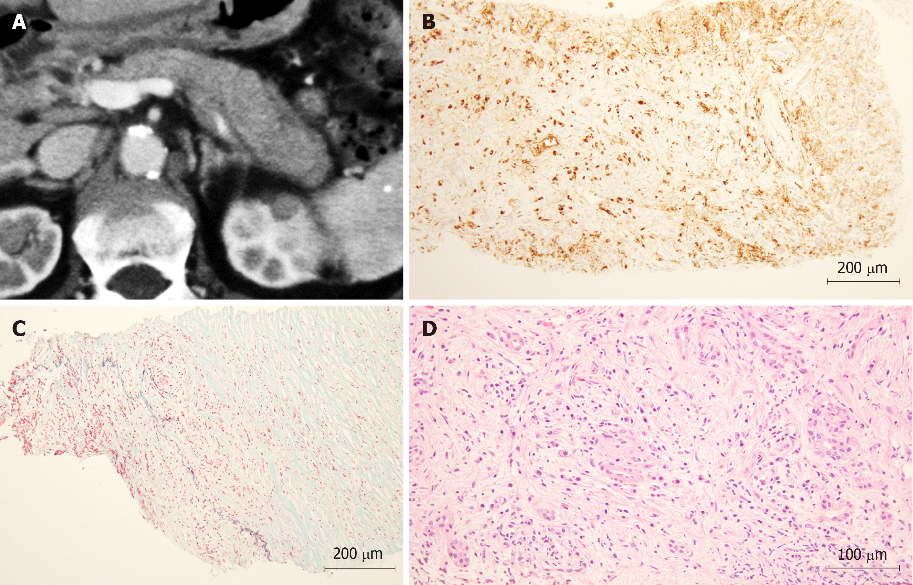
Figure 3 The features of a patient with autoimmune pancreatitis diagnosed by endoscopic ultrasonography-guided fine-needle aspiration with a wet suction technique.
A: The pancreatic tail is swollen with a capsule-like rim sign as observed on abdominal computed tomography; B: Specimen acquired by wet suction technique (WEST) endoscopic ultrasonography-guided fine-needle aspiration (EUS-FNA) (HE × 200): IgG4-positive plasma cells were observed; C: Specimen acquired by WEST EUS-FNA (EM × 200): obliterative phlebitis was observed; D: Specimen acquired by WEST EUS-FNA (HE × 400): fibrosis with a storiform pattern of plasma cells was observed.
- Citation: Sugimoto M, Takagi T, Suzuki R, Konno N, Asama H, Sato Y, Irie H, Watanabe K, Nakamura J, Kikuchi H, Takasumi M, Hashimoto M, Kato T, Hikichi T, Notohara K, Ohira H. Can the wet suction technique change the efficacy of endoscopic ultrasound-guided fine-needle aspiration for diagnosing autoimmune pancreatitis type 1? A prospective single-arm study. World J Clin Cases 2020; 8(1): 88-96
- URL: https://www.wjgnet.com/2307-8960/full/v8/i1/88.htm
- DOI: https://dx.doi.org/10.12998/wjcc.v8.i1.88


