Copyright
©The Author(s) 2020.
World J Clin Cases. Jan 6, 2020; 8(1): 234-241
Published online Jan 6, 2020. doi: 10.12998/wjcc.v8.i1.234
Published online Jan 6, 2020. doi: 10.12998/wjcc.v8.i1.234
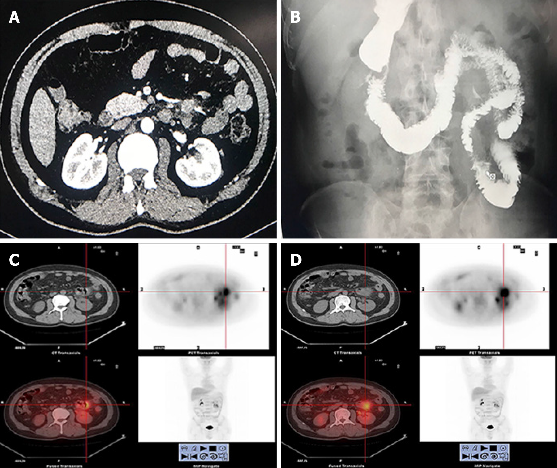
Figure 2 Computed tomography, digestive tract imaging and positron emission tomography/computed tomography results.
A: Local changes at the ascending colon wall; B: Local wall rigidity and mucosal destruction are seen at the proximal jejunum; C, D: Positron emission tomography/computed tomography showing a non-uniform increase in 18-F-fluoro-deoxy-D-glucose metabolism and a space-occupying mass in the small intestine.
- Citation: Dong BL, Dong XH, Zhao HQ, Gao P, Yang XJ. Primary intestinal extranodal natural killer/T-cell lymphoma, nasal type: A case report. World J Clin Cases 2020; 8(1): 234-241
- URL: https://www.wjgnet.com/2307-8960/full/v8/i1/234.htm
- DOI: https://dx.doi.org/10.12998/wjcc.v8.i1.234


