Copyright
©The Author(s) 2020.
World J Clin Cases. Jan 6, 2020; 8(1): 11-19
Published online Jan 6, 2020. doi: 10.12998/wjcc.v8.i1.11
Published online Jan 6, 2020. doi: 10.12998/wjcc.v8.i1.11
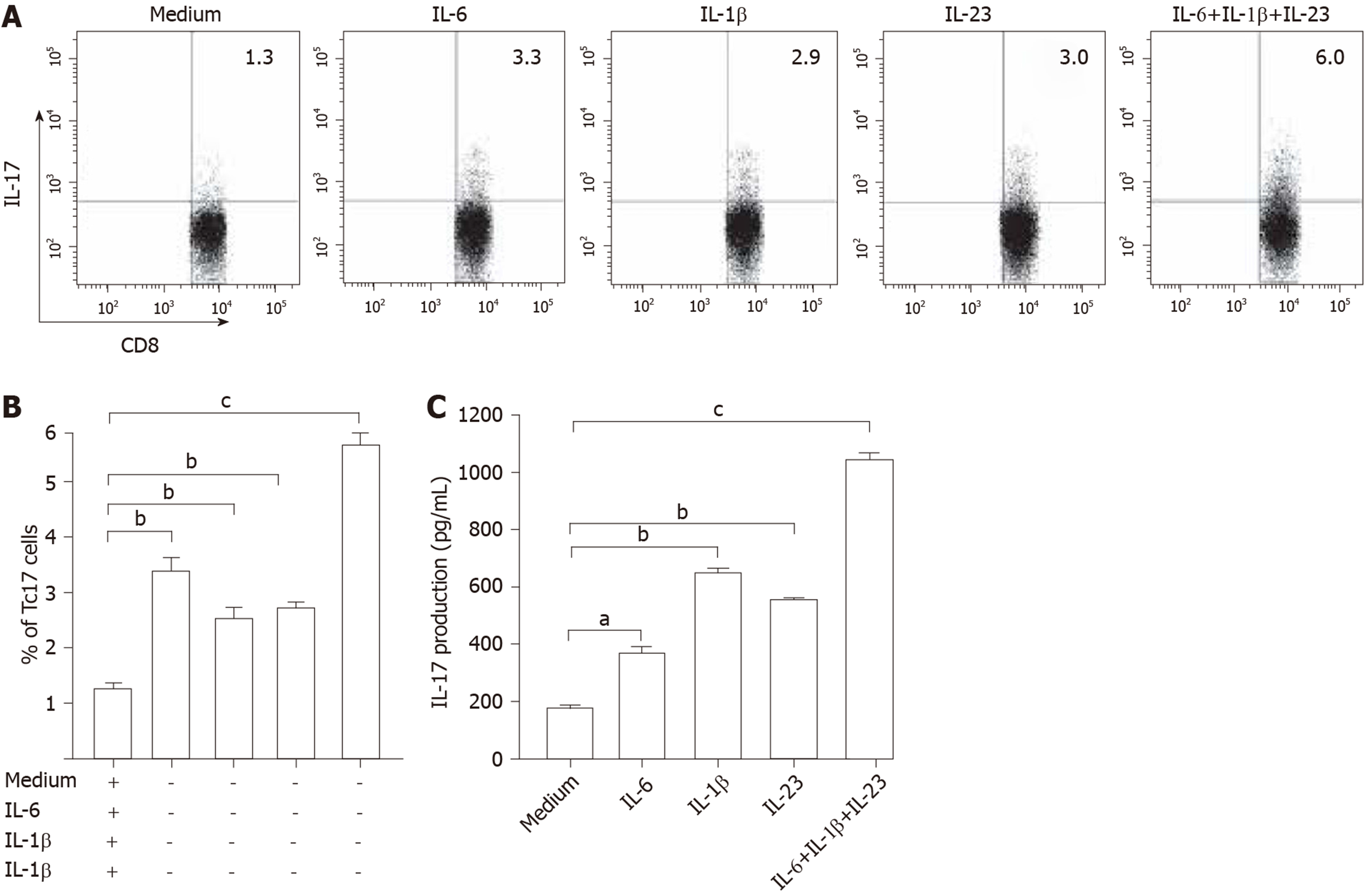
Figure 3 Interleukin-6, interleukin-1β, and interleukin-23 induced differentiation of Tc17 cells.
A-C: Peripheral CD8+ T cells and blood monocytes were co-cultured as described in Materials and Methods. Representative data and statistical analysis of Tc17 cell percentage in CD8+ T cells and interleukin-17 production in the culture supernatants from the co-culture systems. Comparisons were performed using the t-test. aP < 0.05, bP < 0.01, cP < 0.001. Error bars represent SE. IL: Interleukin.
- Citation: Zhang ZS, Gu Y, Liu BG, Tang H, Hua Y, Wang J. Oncogenic role of Tc17 cells in cervical cancer development. World J Clin Cases 2020; 8(1): 11-19
- URL: https://www.wjgnet.com/2307-8960/full/v8/i1/11.htm
- DOI: https://dx.doi.org/10.12998/wjcc.v8.i1.11


