Copyright
©The Author(s) 2019.
World J Clin Cases. May 6, 2019; 7(9): 1066-1072
Published online May 6, 2019. doi: 10.12998/wjcc.v7.i9.1066
Published online May 6, 2019. doi: 10.12998/wjcc.v7.i9.1066
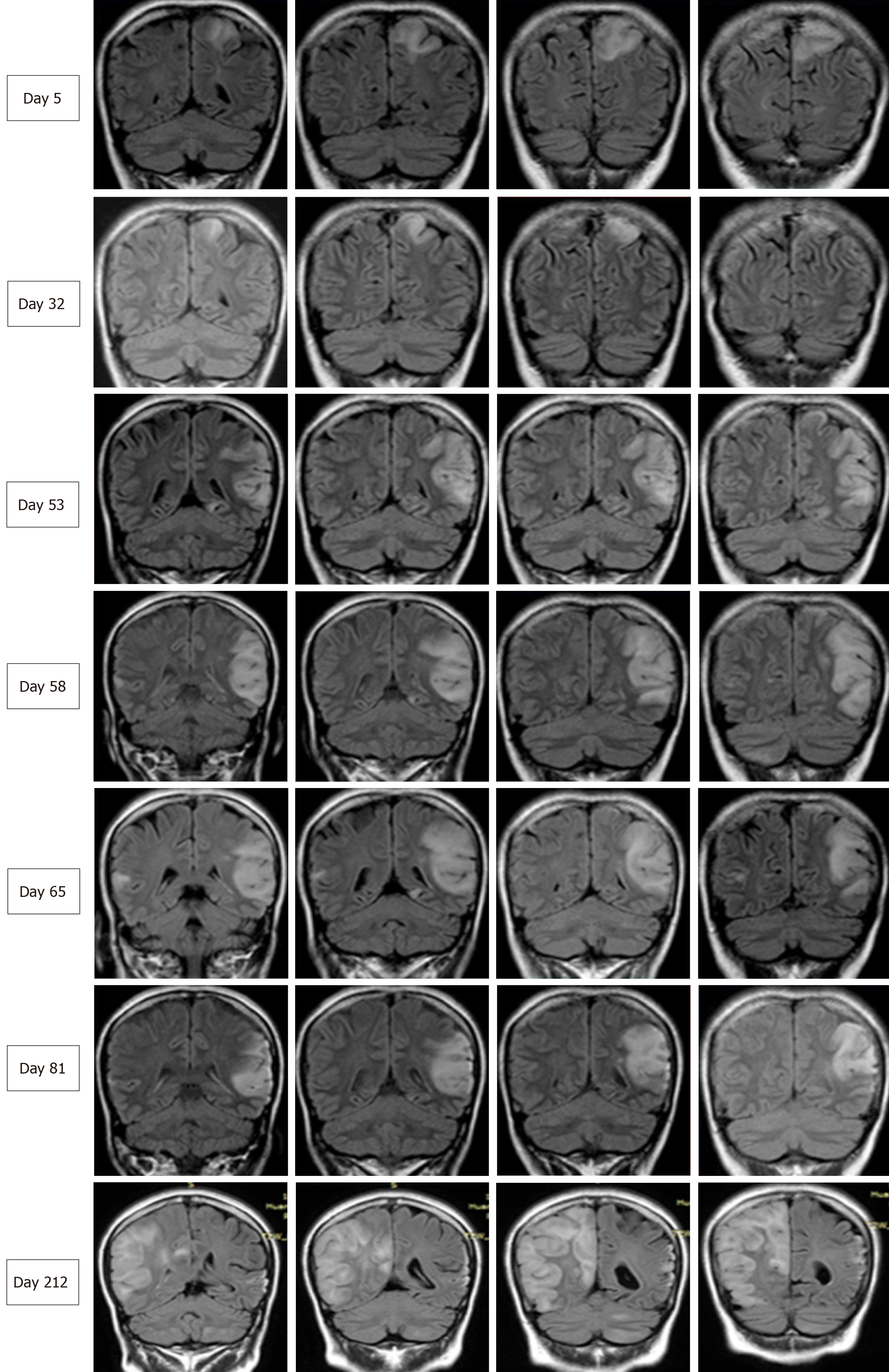
Figure 2 Serial brain images covering three recurrent stroke-like episodes in the present patient over a 7-mo period.
Four representative coronal slices were serially shown in chronological order. Fluid-attenuated inversion recovery images on days 5, 32, 58, 65, 81, and 212 revealed hyperintensity signals appearing recurrently in various cortical and subcortical areas, mainly in the parietal and temporal lobes, and the progressive development of cortical atrophy.
- Citation: Fu XL, Zhou XX, Shi Z, Zheng WC. Adult-onset mitochondrial encephalopathy in association with the MT-ND3 T10158C mutation exhibits unique characteristics: A case report. World J Clin Cases 2019; 7(9): 1066-1072
- URL: https://www.wjgnet.com/2307-8960/full/v7/i9/1066.htm
- DOI: https://dx.doi.org/10.12998/wjcc.v7.i9.1066


