Copyright
©The Author(s) 2019.
World J Clin Cases. May 6, 2019; 7(9): 1053-1059
Published online May 6, 2019. doi: 10.12998/wjcc.v7.i9.1053
Published online May 6, 2019. doi: 10.12998/wjcc.v7.i9.1053
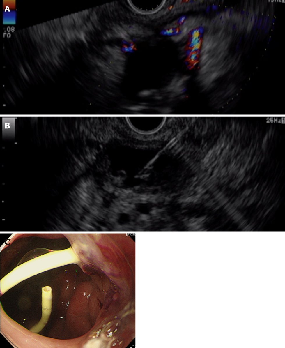
Figure 4 Endoscopic ultrasound-guided drainage.
A: A linear array echo-endoscope shows many vessels surrounding the pancreatic fluid collection and stomach; B, C: The pancreatic fluid collection is punctured using a 19-gauge needle, carefully avoiding the vessels (B), and a double pigtail catheter (6 French/4 cm) is inserted transmurally (C).
- Citation: Yamada R, Umeda Y, Shiono Y, Okuse H, Kuroda N, Tsuboi J, Inoue H, Hamada Y, Tanaka K, Horiki N, Takei Y. Management of the late effects of disconnected pancreatic duct syndrome: A case report. World J Clin Cases 2019; 7(9): 1053-1059
- URL: https://www.wjgnet.com/2307-8960/full/v7/i9/1053.htm
- DOI: https://dx.doi.org/10.12998/wjcc.v7.i9.1053


