Copyright
©The Author(s) 2019.
World J Clin Cases. Apr 26, 2019; 7(8): 1001-1005
Published online Apr 26, 2019. doi: 10.12998/wjcc.v7.i8.1001
Published online Apr 26, 2019. doi: 10.12998/wjcc.v7.i8.1001
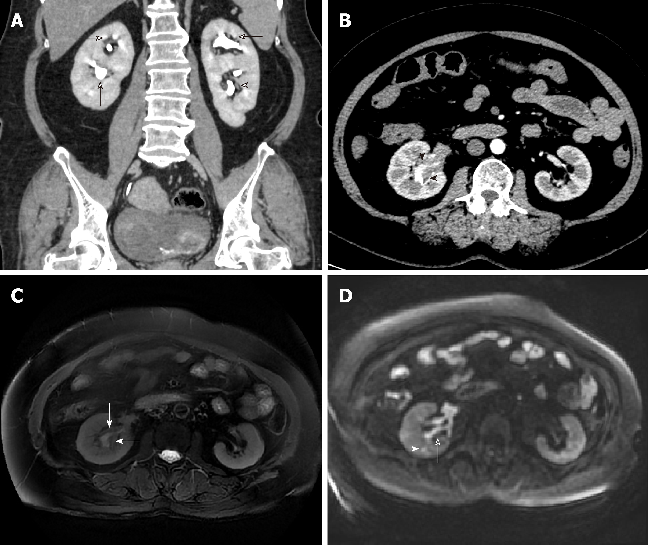
Figure 1 Plasma cell type of Castleman’s disease involving the renal sinus and adjacent parenchyma in a 65-year-old woman.
A: Coronal contrast-enhanced computed tomography (CT) image showing bilateral renal pelvis duplication; B: Axial contrast-enhanced CT image obtained at the renal hilum level shows mildly homogenous enhancement of a soft tissue mass in the right lower renal sinus; C: T2-weighted images (TR/TE, 10000/82.6) showing a soft tissue mass in the right renal sinus with slightly hypointense signals compared to that of the renal cortex; D: Diffusion-weighted images (b = 800 s/mm2) showing that the mass (open arrow) is mildly hyperintense, and a hyperintensity lesion is in the renal parenchyma (solid arrow).
- Citation: Guo XW, Jia XD, Shen SS, Ji H, Chen YM, Du Q, Zhang SQ. Radiologic features of Castleman’s disease involving the renal sinus: A case report and review of the literature. World J Clin Cases 2019; 7(8): 1001-1005
- URL: https://www.wjgnet.com/2307-8960/full/v7/i8/1001.htm
- DOI: https://dx.doi.org/10.12998/wjcc.v7.i8.1001


