Copyright
©The Author(s) 2019.
World J Clin Cases. Mar 26, 2019; 7(6): 717-726
Published online Mar 26, 2019. doi: 10.12998/wjcc.v7.i6.717
Published online Mar 26, 2019. doi: 10.12998/wjcc.v7.i6.717
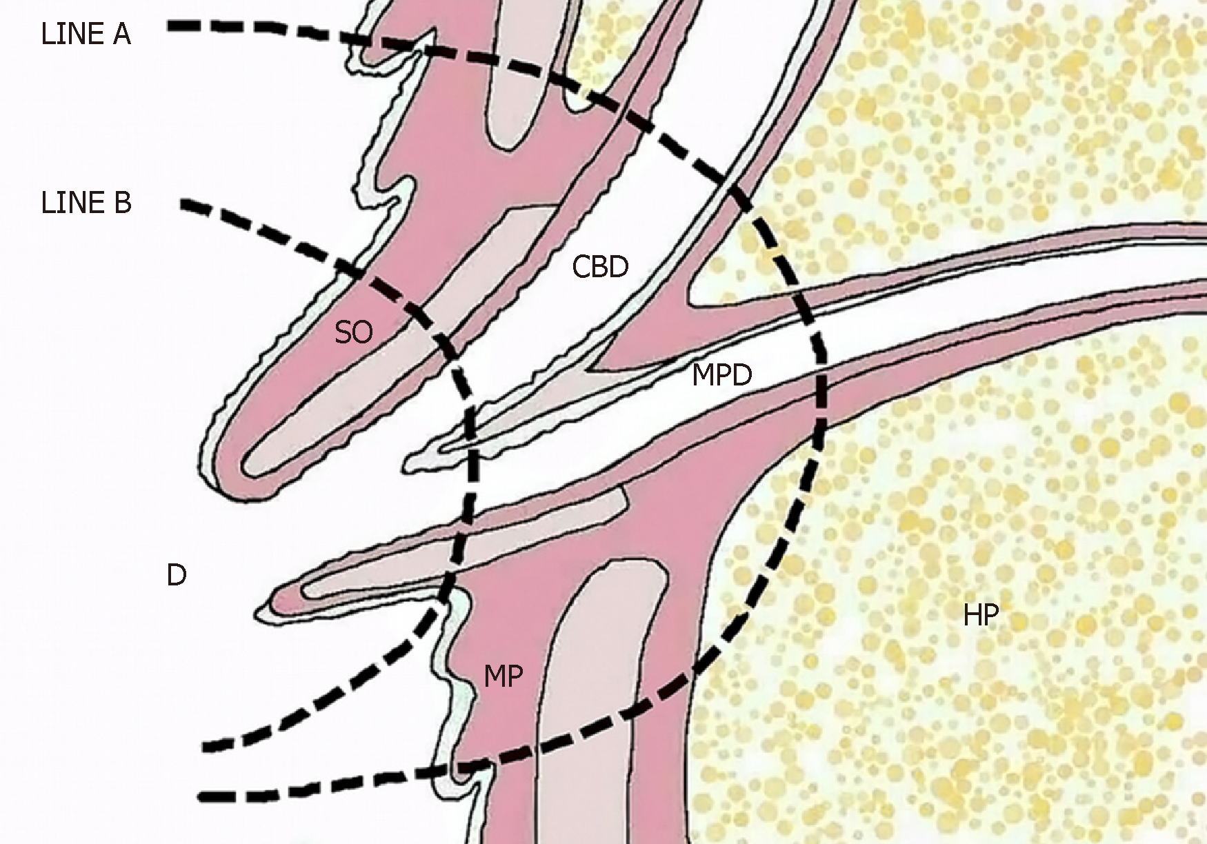
Figure 3 Diagrammatic drawing showing the ampulla of Vater.
LINE A: Local resection line; LINE B: Endoscopic papillectomy line prior to the muscularis propria. CBD: Common bile duct; MP: Muscularis propria; HP: Head of the pancreas; MPD: Main pancreatic duct; SO: Sphincter of Oddi; D: Duodenum.
- Citation: Liu F, Cheng JL, Cui J, Xu ZZ, Fu Z, Liu J, Tian H. Surgical method choice and coincidence rate of pathological diagnoses in transduodenal ampullectomy: A retrospective case series study and review of the literature. World J Clin Cases 2019; 7(6): 717-726
- URL: https://www.wjgnet.com/2307-8960/full/v7/i6/717.htm
- DOI: https://dx.doi.org/10.12998/wjcc.v7.i6.717


