Copyright
©The Author(s) 2019.
World J Clin Cases. Mar 26, 2019; 7(6): 717-726
Published online Mar 26, 2019. doi: 10.12998/wjcc.v7.i6.717
Published online Mar 26, 2019. doi: 10.12998/wjcc.v7.i6.717
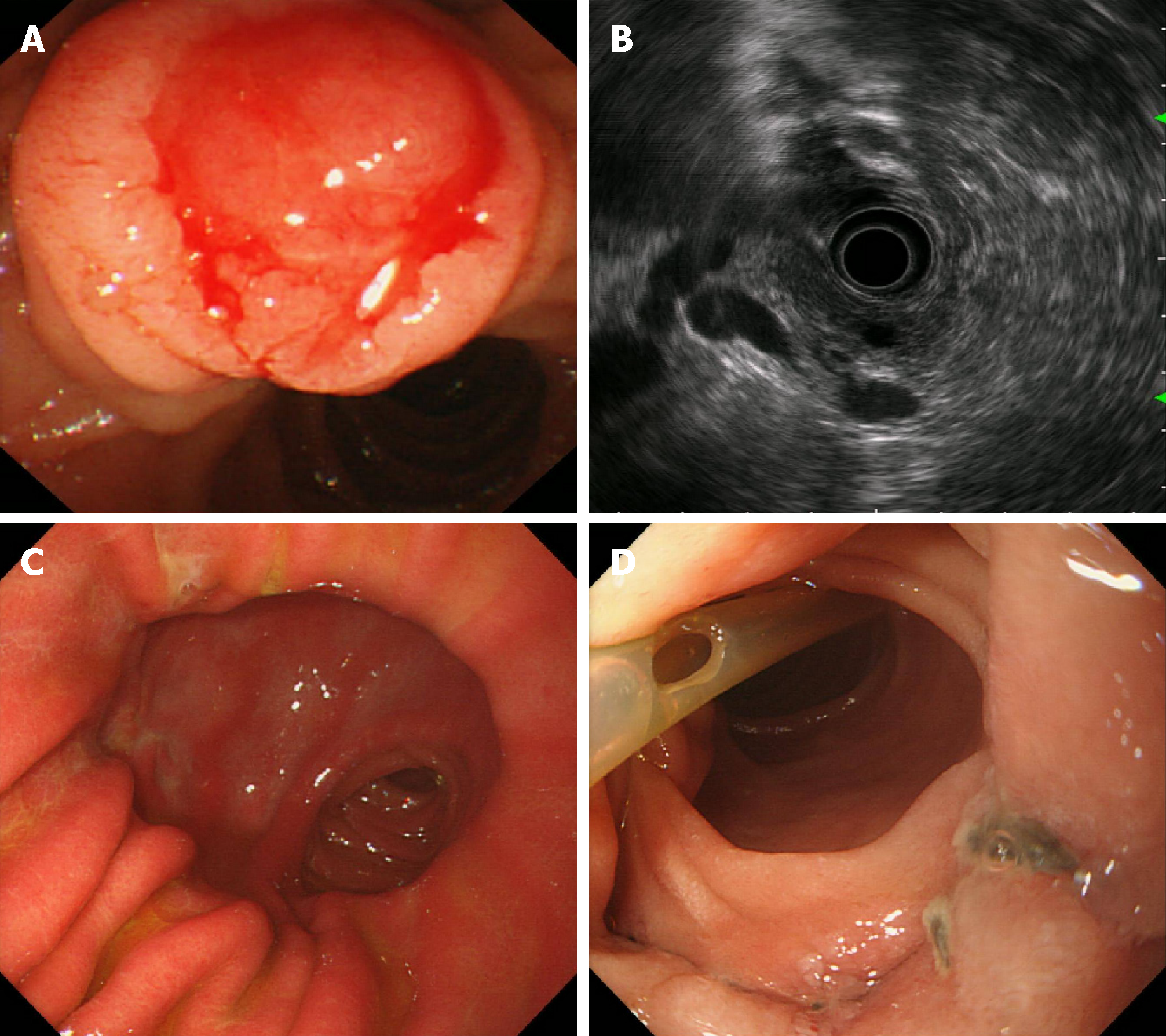
Figure 1 Gastroscopic images and endoscopic ultrasonography.
A: A swollen duodenal papilla that bled easily when touched; B: Periampullary endoscopic ultrasound showing bile duct and pancreatic duct expansion; C: Gastroscopic image 3 mo after operation; a small amount of bile refluxed to the stomach cavity, with no gastric ulcer; D: Site of bile duct, pancreatic duct, and duodenal anastomosis replantation; no anastomotic tumor relapse; visible suture knot; pancreatic duct supporting tube fell off.
- Citation: Liu F, Cheng JL, Cui J, Xu ZZ, Fu Z, Liu J, Tian H. Surgical method choice and coincidence rate of pathological diagnoses in transduodenal ampullectomy: A retrospective case series study and review of the literature. World J Clin Cases 2019; 7(6): 717-726
- URL: https://www.wjgnet.com/2307-8960/full/v7/i6/717.htm
- DOI: https://dx.doi.org/10.12998/wjcc.v7.i6.717


