
















©The Author(s) 2019.
World J Clin Cases. Feb 26, 2019; 7(4): 538-547
Published online Feb 26, 2019. doi: 10.12998/wjcc.v7.i4.538
Published online Feb 26, 2019. doi: 10.12998/wjcc.v7.i4.538
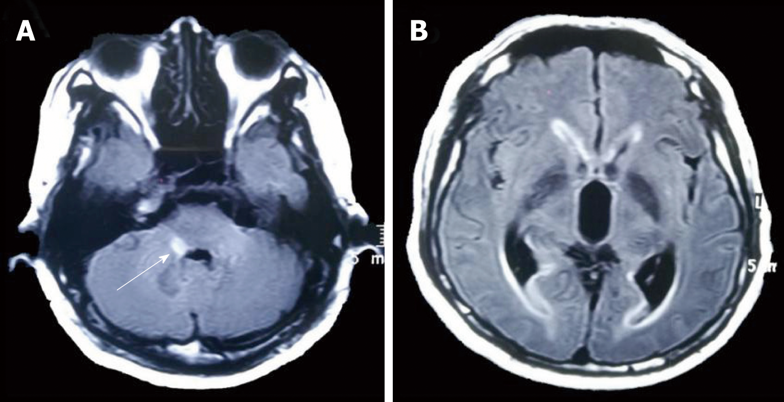
Figure 1 Axial brian T2-FLAIR magnetic resonance imaging shows a hyperintense lesion of the right pons (A, white arrow), and prominent temporal horns with enlargement of ventricles (B) on the 4th d of administration.
- Citation: Liang JJ, He XY, Ye H. Rhombencephalitis caused by Listeria monocytogenes with hydrocephalus and intracranial hemorrhage: A case report and review of the literature. World J Clin Cases 2019; 7(4): 538-547
- URL: https://www.wjgnet.com/2307-8960/full/v7/i4/538.htm
- DOI: https://dx.doi.org/10.12998/wjcc.v7.i4.538