Copyright
©The Author(s) 2019.
World J Clin Cases. Feb 26, 2019; 7(4): 473-481
Published online Feb 26, 2019. doi: 10.12998/wjcc.v7.i4.473
Published online Feb 26, 2019. doi: 10.12998/wjcc.v7.i4.473
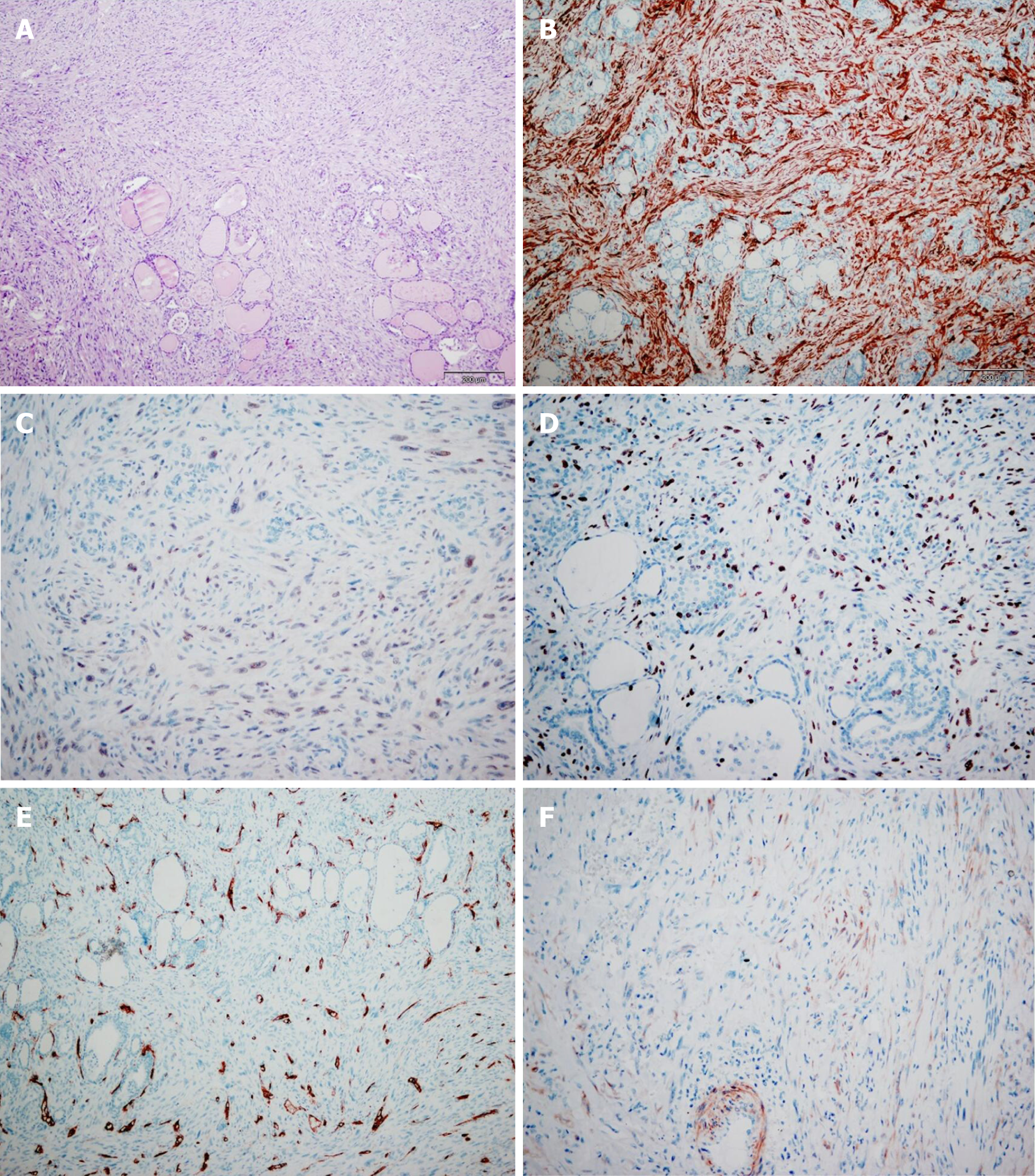
Figure 1 The tumor cells tested positive for alpha smooth muscle actin, calponin, H-caldesmon, but were negative for CD34, p53, estrogen receptor, progesterone receptor, and Epstein-Barr virus.
In approximately 25% of all the tumor cells, the Ki67 proliferative index was positive. A: Pathological finding of thyroid gland stained (10 ×); B: Positive immunohistochemical staining for α-smooth muscle actin (10 ×); C: Negative immunohistochemical staining for P53 (20 ×); D: Pathological finding of 25% positive Ki67 index in tumor cells (20 ×); E: Negative immunohistochemical staining for CD34 (10 ×); F: Positive immunohistochemical staining for calponin (20 ×).
- Citation: Vujosevic S, Krnjevic D, Bogojevic M, Vuckovic L, Filipovic A, Dunđerović D, Sopta J. Primary leiomyosarcoma of the thyroid gland with prior malignancy and radiotherapy: A case report and review of literature. World J Clin Cases 2019; 7(4): 473-481
- URL: https://www.wjgnet.com/2307-8960/full/v7/i4/473.htm
- DOI: https://dx.doi.org/10.12998/wjcc.v7.i4.473


