Copyright
©The Author(s) 2019.
World J Clin Cases. Feb 26, 2019; 7(4): 441-451
Published online Feb 26, 2019. doi: 10.12998/wjcc.v7.i4.441
Published online Feb 26, 2019. doi: 10.12998/wjcc.v7.i4.441
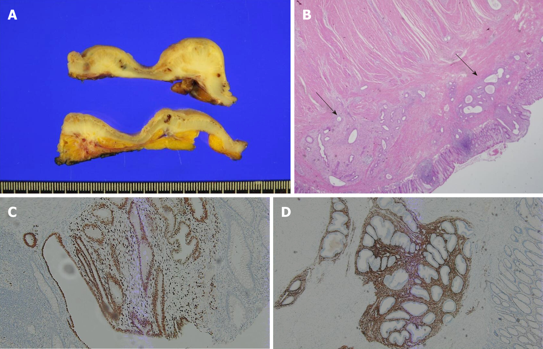
Figure 4 Pathologic and histologic findings.
A: Gross specimen sections showing endometriotic nodules infiltrating from the outer layers; B: Endometrial gland (arrow) in the submucosal layer, infiltrating to the muscularis mucosa (hematoxylin and eosin stain); C: Immunohistochemical examination for endometrial gland expressing ER; D: Immunohistochemical examination for the stroma expressing CD10.
- Citation: Bong JW, Yu CS, Lee JL, Kim CW, Yoon YS, Park IJ, Lim SB, Kim JC. Intestinal endometriosis: Diagnostic ambiguities and surgical outcomes. World J Clin Cases 2019; 7(4): 441-451
- URL: https://www.wjgnet.com/2307-8960/full/v7/i4/441.htm
- DOI: https://dx.doi.org/10.12998/wjcc.v7.i4.441


