Copyright
©The Author(s) 2019.
World J Clin Cases. Dec 26, 2019; 7(24): 4391-4397
Published online Dec 26, 2019. doi: 10.12998/wjcc.v7.i24.4391
Published online Dec 26, 2019. doi: 10.12998/wjcc.v7.i24.4391
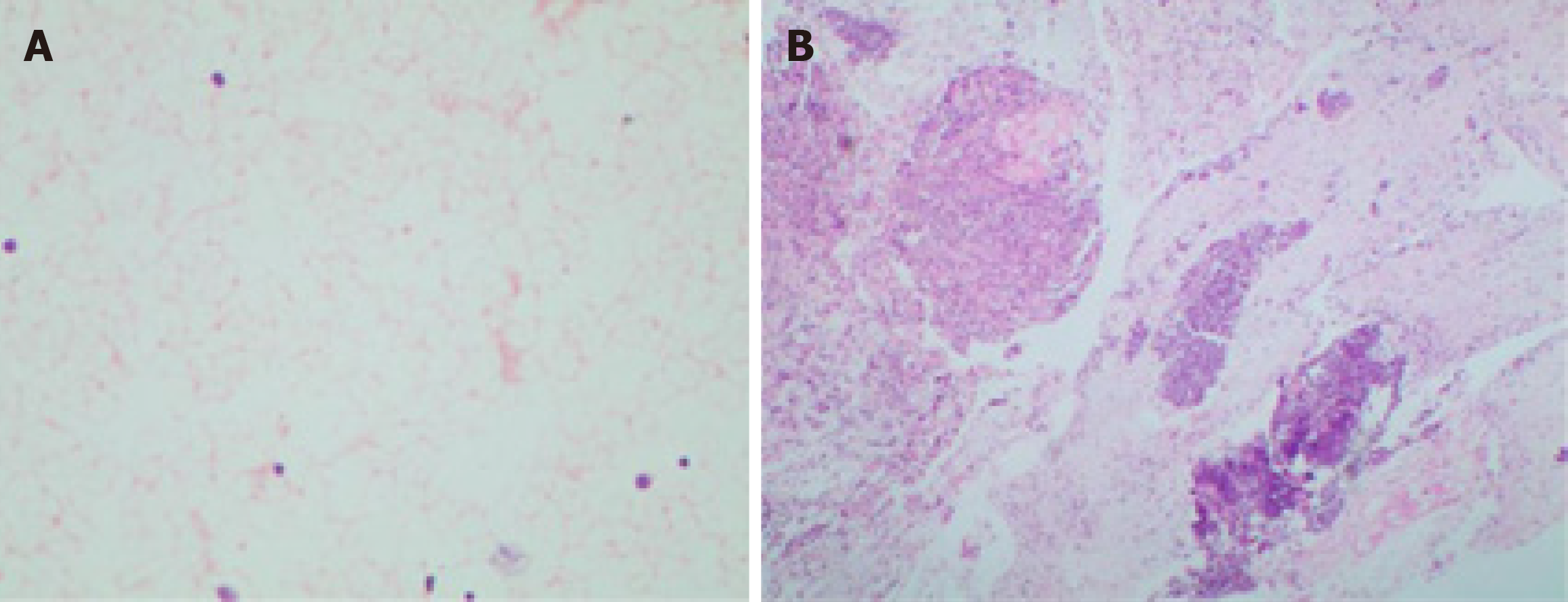
Figure 5 Hematoxylin eosin staining of the smear and tissue.
A: Hematoxylin eosin staining of the smear: Scattered lymphocytes with normal morphology; B: Hematoxylin eosin staining of the tissue: A large number of lymphocytes with normal morphology, dominated by B cells.
- Citation: Xu XY, Liu XQ, Du HW, Liu JH. Castleman disease in the hepatic-gastric space: A case report. World J Clin Cases 2019; 7(24): 4391-4397
- URL: https://www.wjgnet.com/2307-8960/full/v7/i24/4391.htm
- DOI: https://dx.doi.org/10.12998/wjcc.v7.i24.4391


