Copyright
©The Author(s) 2019.
World J Clin Cases. Dec 26, 2019; 7(24): 4377-4383
Published online Dec 26, 2019. doi: 10.12998/wjcc.v7.i24.4377
Published online Dec 26, 2019. doi: 10.12998/wjcc.v7.i24.4377
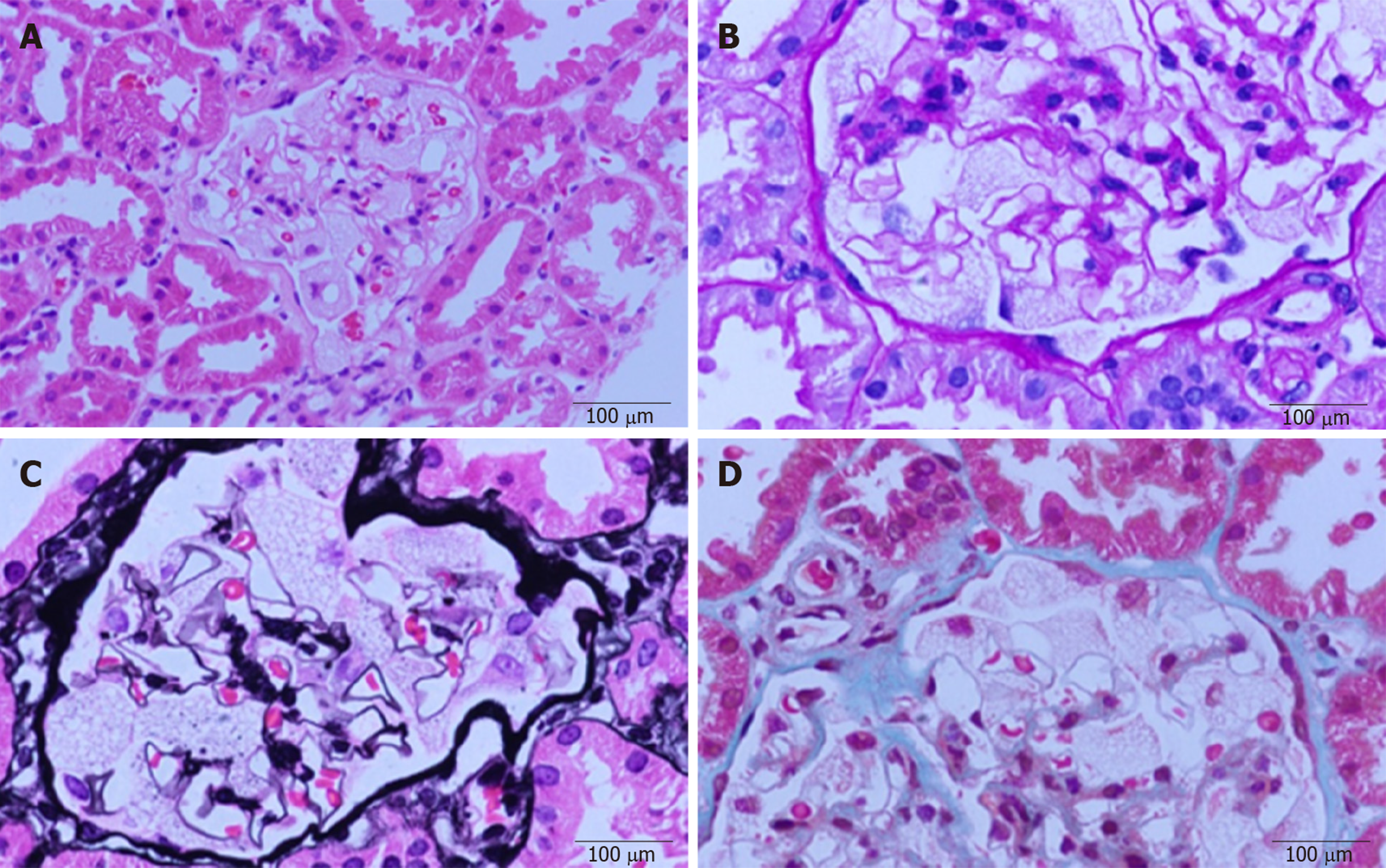
Figure 1 Light microscopic images.
Diffuse enlargement and vacuolar degeneration of glomerular visceral epithelial cells are seen. A: Hematoxylin-eosin staining; B: Periodic acid-Schiff staining; C: Periodic acid-silver methenamine staining; and D: Masson staining.
- Citation: Wu SZ, Liang X, Geng J, Zhang MB, Xie N, Su XY. Hydroxychloroquine-induced renal phospholipidosis resembling Fabry disease in undifferentiated connective tissue disease: A case report. World J Clin Cases 2019; 7(24): 4377-4383
- URL: https://www.wjgnet.com/2307-8960/full/v7/i24/4377.htm
- DOI: https://dx.doi.org/10.12998/wjcc.v7.i24.4377


