
















©The Author(s) 2019.
World J Clin Cases. Dec 26, 2019; 7(24): 4334-4341
Published online Dec 26, 2019. doi: 10.12998/wjcc.v7.i24.4334
Published online Dec 26, 2019. doi: 10.12998/wjcc.v7.i24.4334
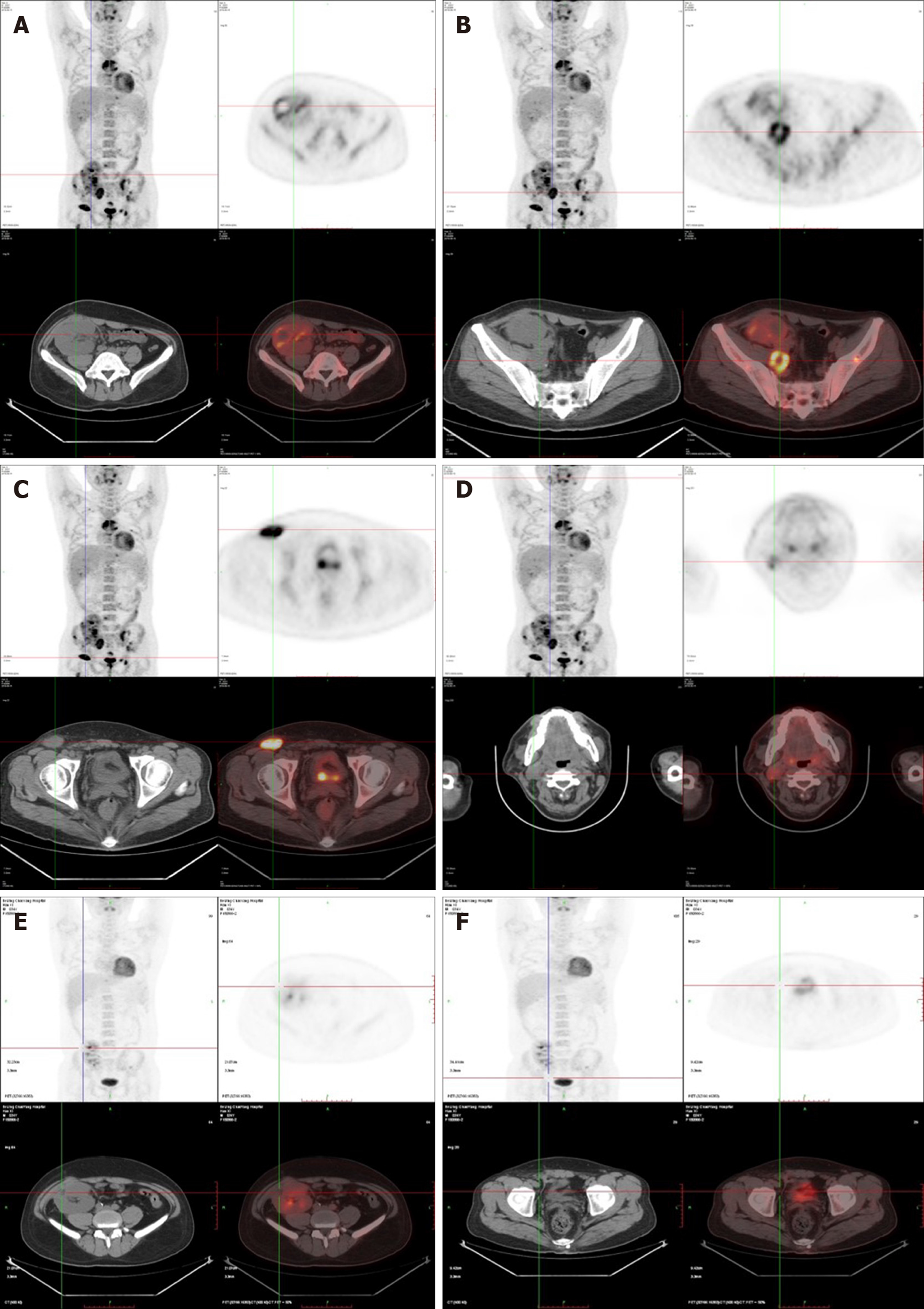
Figure 3 Positron emission tomography computed tomography.
A-D: Prior to treatment: Lymphadenectasis and increased bone metabolism in the neck, abdominal cavity, retroperitoneum, and inguinal region. The transplanted kidney was invaded, which was accompanied by necrotic lesions; E, F: After treatment: No increased metabolism in multiple lymph nodes in the neck, abdominal cavity, retroperitoneum, and inguinal region. The number and volume of the abnormal lesions in the transplanted kidney and bone metabolism were significantly reduced.
- Citation: Sun ZJ, Hu XP, Fan BH, Wang W. Epstein-Barr virus-positive post-transplant lymphoproliferative disordepresenting as hematochezia and enterobrosis in renal transplant recipients in China: A report of two cases. World J Clin Cases 2019; 7(24): 4334-4341
- URL: https://www.wjgnet.com/2307-8960/full/v7/i24/4334.htm
- DOI: https://dx.doi.org/10.12998/wjcc.v7.i24.4334