Copyright
©The Author(s) 2019.
World J Clin Cases. Dec 26, 2019; 7(24): 4299-4306
Published online Dec 26, 2019. doi: 10.12998/wjcc.v7.i24.4299
Published online Dec 26, 2019. doi: 10.12998/wjcc.v7.i24.4299
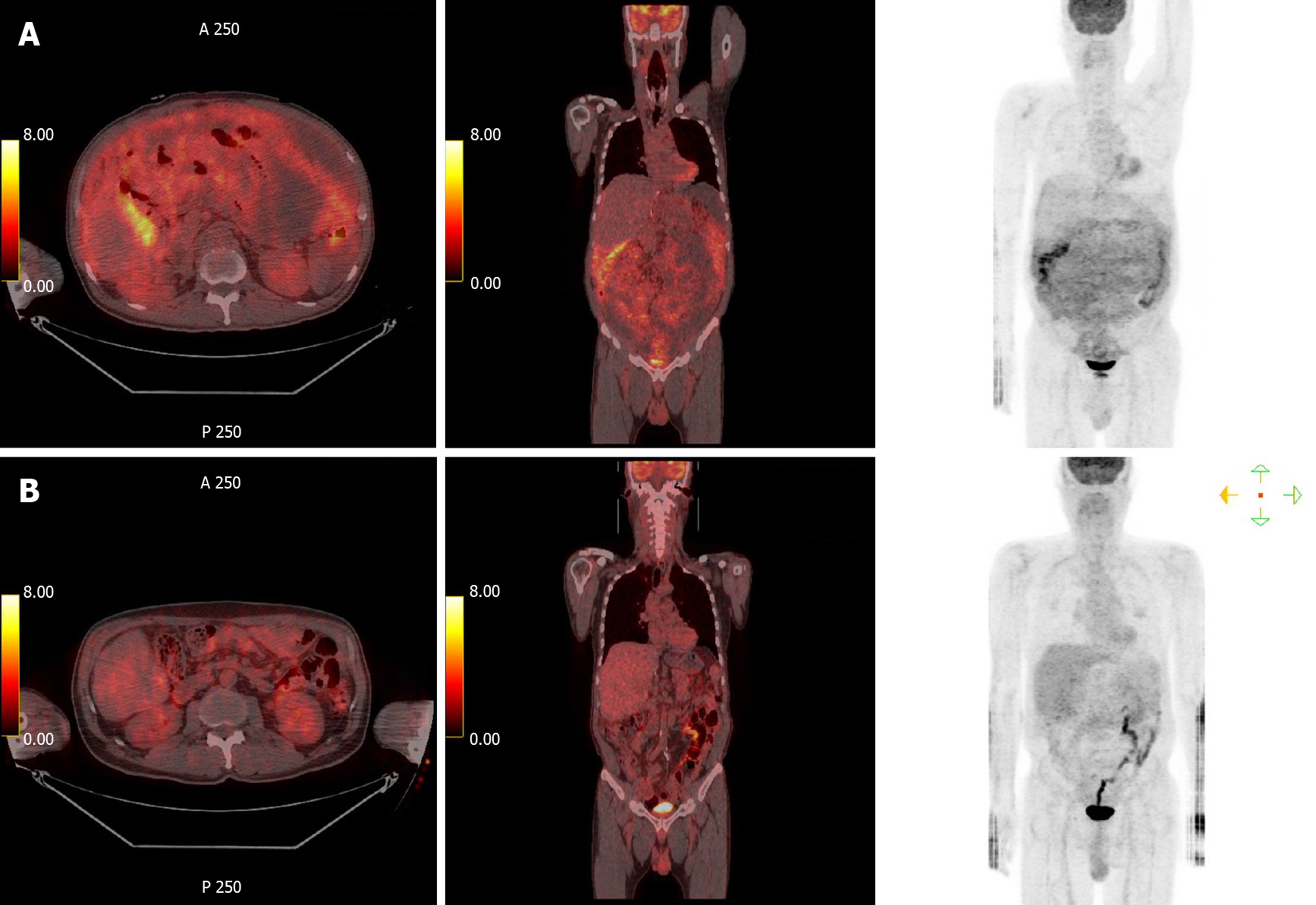
Figure 2 Positron emission tomography-computed tomography.
A: Positron emission tomography (PET)-computed tomography (CT) did not depict abnormal hypermetabolism in solid organs, lymph nodes, or digestive organs, but it did depict diffuse hypermetabolism in the peritoneum and omentum; B: Post-chemotherapy PET-CT depicted loss of diffuse hypermetabolic lesion in the peritoneum.
- Citation: Kim HB, Hong R, Na YS, Choi WY, Park SG, Lee HJ. Isolated peritoneal lymphomatosis defined as post-transplant lymphoproliferative disorder after a liver transplant: A case report. World J Clin Cases 2019; 7(24): 4299-4306
- URL: https://www.wjgnet.com/2307-8960/full/v7/i24/4299.htm
- DOI: https://dx.doi.org/10.12998/wjcc.v7.i24.4299


