Copyright
©The Author(s) 2019.
World J Clin Cases. Dec 26, 2019; 7(24): 4196-4207
Published online Dec 26, 2019. doi: 10.12998/wjcc.v7.i24.4196
Published online Dec 26, 2019. doi: 10.12998/wjcc.v7.i24.4196
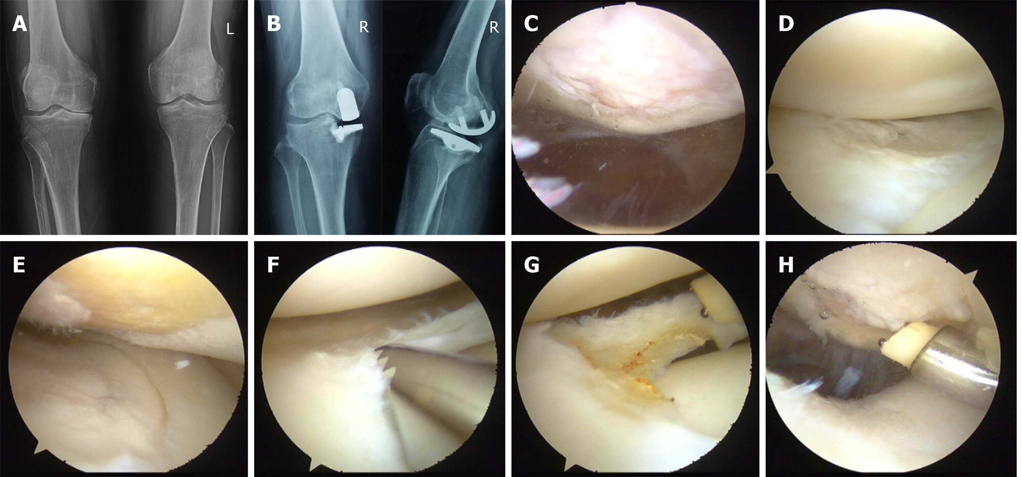
Figure 5 Surgical procedure and images of a 53-year-old woman.
A: Preoperative radiographs showing unicompartmental osteoarthritis on the right knee; B: Image taken at 8 years after arthroscopy combined with unicondylar knee arthroplasty showing that the prosthesis is in good position and the patient is experiencing good recovery as well as normal knee mobility. C: IV-degree injury of patellofemoral articular cartilage; D: Normal cartilage and meniscus injury of lateral compartment; E: IV-degree injury of internal femoral cartilage; F and G: Trimming the lateral meniscus injury site; H: Trimming the wound of patellofemoral articular cartilage.
- Citation: Wang HR, Li ZL, Li J, Wang YX, Zhao ZD, Li W. Arthroscopy combined with unicondylar knee arthroplasty for treatment of isolated unicompartmental knee arthritis: A long-term comparison. World J Clin Cases 2019; 7(24): 4196-4207
- URL: https://www.wjgnet.com/2307-8960/full/v7/i24/4196.htm
- DOI: https://dx.doi.org/10.12998/wjcc.v7.i24.4196


