
















©The Author(s) 2019.
World J Clin Cases. Dec 6, 2019; 7(23): 4150-4156
Published online Dec 6, 2019. doi: 10.12998/wjcc.v7.i23.4150
Published online Dec 6, 2019. doi: 10.12998/wjcc.v7.i23.4150
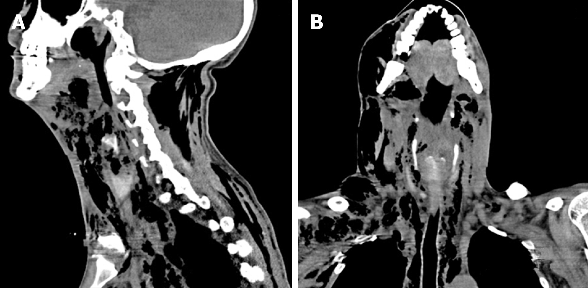
Figure 1 Sagittal and coronal computed tomography scans of the maxillofacial, neck and chest regions.
Sagittal (A) and coronal (B) computed tomography scans of the maxillofacial, neck and chest regions showing extensive swelling and pneumatosis in soft tissues of bilateral oral, maxillofacial, temporal, cervical, parapharyngeal space, mediastinum, chest and back regions.
- Citation: Dai TG, Ran HB, Qiu YX, Xu B, Cheng JQ, Liu YK. Fatal complications in a patient with severe multi-space infections in the oral and maxillofacial head and neck regions: A case report. World J Clin Cases 2019; 7(23): 4150-4156
- URL: https://www.wjgnet.com/2307-8960/full/v7/i23/4150.htm
- DOI: https://dx.doi.org/10.12998/wjcc.v7.i23.4150