Copyright
©The Author(s) 2019.
World J Clin Cases. Dec 6, 2019; 7(23): 4036-4043
Published online Dec 6, 2019. doi: 10.12998/wjcc.v7.i23.4036
Published online Dec 6, 2019. doi: 10.12998/wjcc.v7.i23.4036
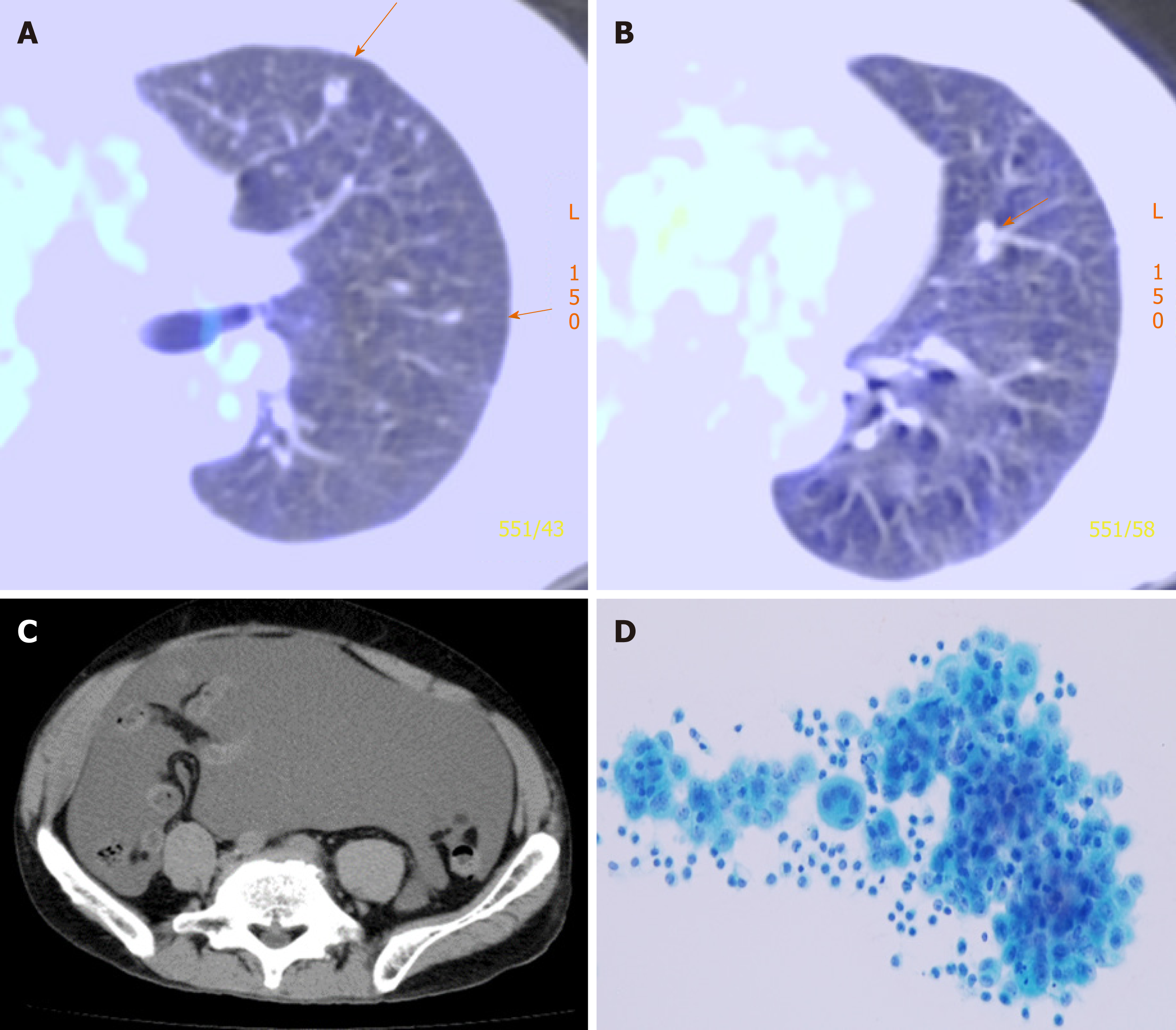
Figure 1 Postoperative recurrence of malignant mesothelioma.
A, B: F-18 fluorodeoxyglucose positron emission tomography/computed tomography (CT) showing multiple pulmonary metastases (orange arrows) that occurred 13 mo postoperatively; C: CT showing massive ascites after eight courses of pemetrexed therapy; D: A photomicrograph of mesothelioma cells in the ascitic fluid obtained via abdominocentesis.
- Citation: Anayama T, Taguchi M, Tatenuma T, Okada H, Miyazaki R, Hirohashi K, Kume M, Matsusaki K, Orihashi K. In-vitro proliferation assay with recycled ascitic cancer cells in malignant pleural mesothelioma: A case report. World J Clin Cases 2019; 7(23): 4036-4043
- URL: https://www.wjgnet.com/2307-8960/full/v7/i23/4036.htm
- DOI: https://dx.doi.org/10.12998/wjcc.v7.i23.4036


