Copyright
©The Author(s) 2019.
World J Clin Cases. Dec 6, 2019; 7(23): 4011-4019
Published online Dec 6, 2019. doi: 10.12998/wjcc.v7.i23.4011
Published online Dec 6, 2019. doi: 10.12998/wjcc.v7.i23.4011
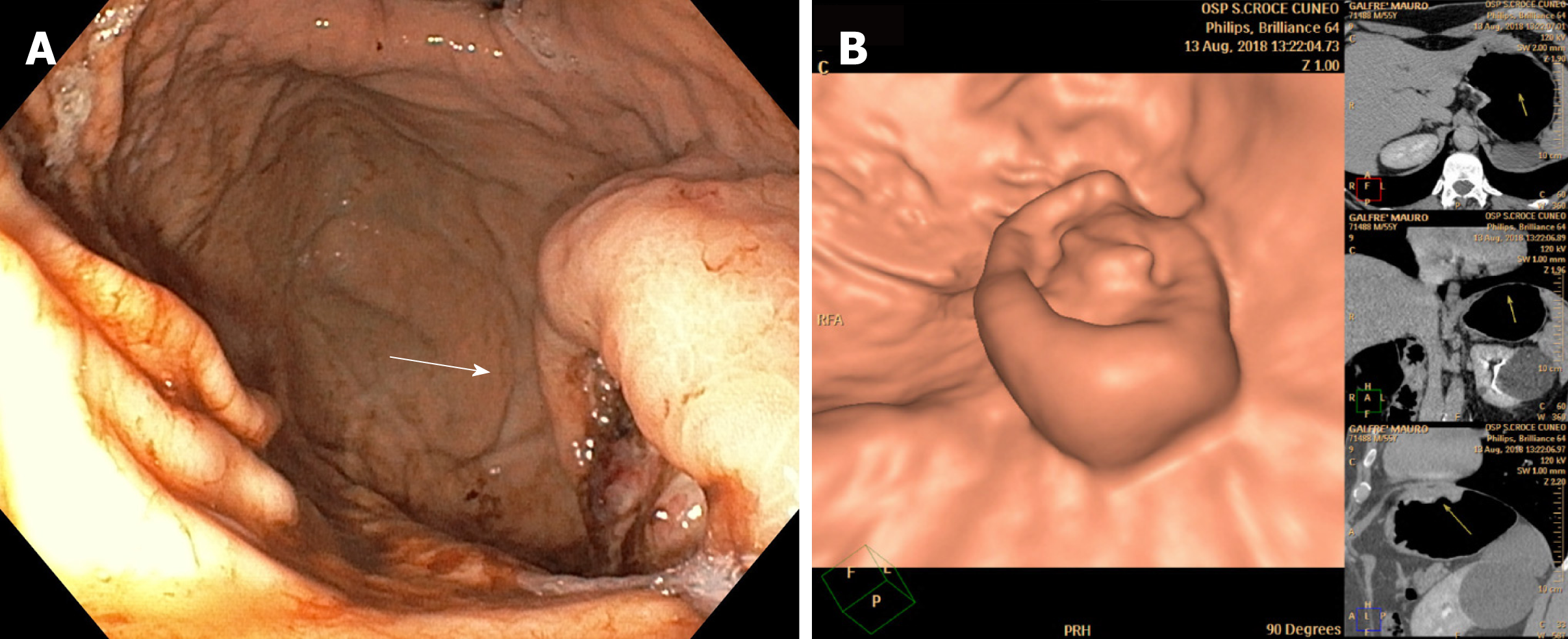
Figure 1 Upper endoscopy and computed tomography gastrography findings at surgery.
A: Upper endoscopy showed an ulcerative lesion of the gastric fundus with spontaneous bleeding; B: Computed tomography gastrography showed a relatively well-defined mass with ulceration of the gastric fundus, 3 cm below the esophago-gastric junction, with heterogeneous enhancement, measuring approximately 60 mm.
- Citation: Marano A, Maione F, Woo Y, Pellegrino L, Geretto P, Sasia D, Fortunato M, Orcioni GF, Priotto R, Fasoli R, Borghi F. Robotic wedge resection of a rare gastric perivascular epithelioid cell tumor: A case report. World J Clin Cases 2019; 7(23): 4011-4019
- URL: https://www.wjgnet.com/2307-8960/full/v7/i23/4011.htm
- DOI: https://dx.doi.org/10.12998/wjcc.v7.i23.4011


