Copyright
©The Author(s) 2019.
World J Clin Cases. Dec 6, 2019; 7(23): 4004-4010
Published online Dec 6, 2019. doi: 10.12998/wjcc.v7.i23.4004
Published online Dec 6, 2019. doi: 10.12998/wjcc.v7.i23.4004
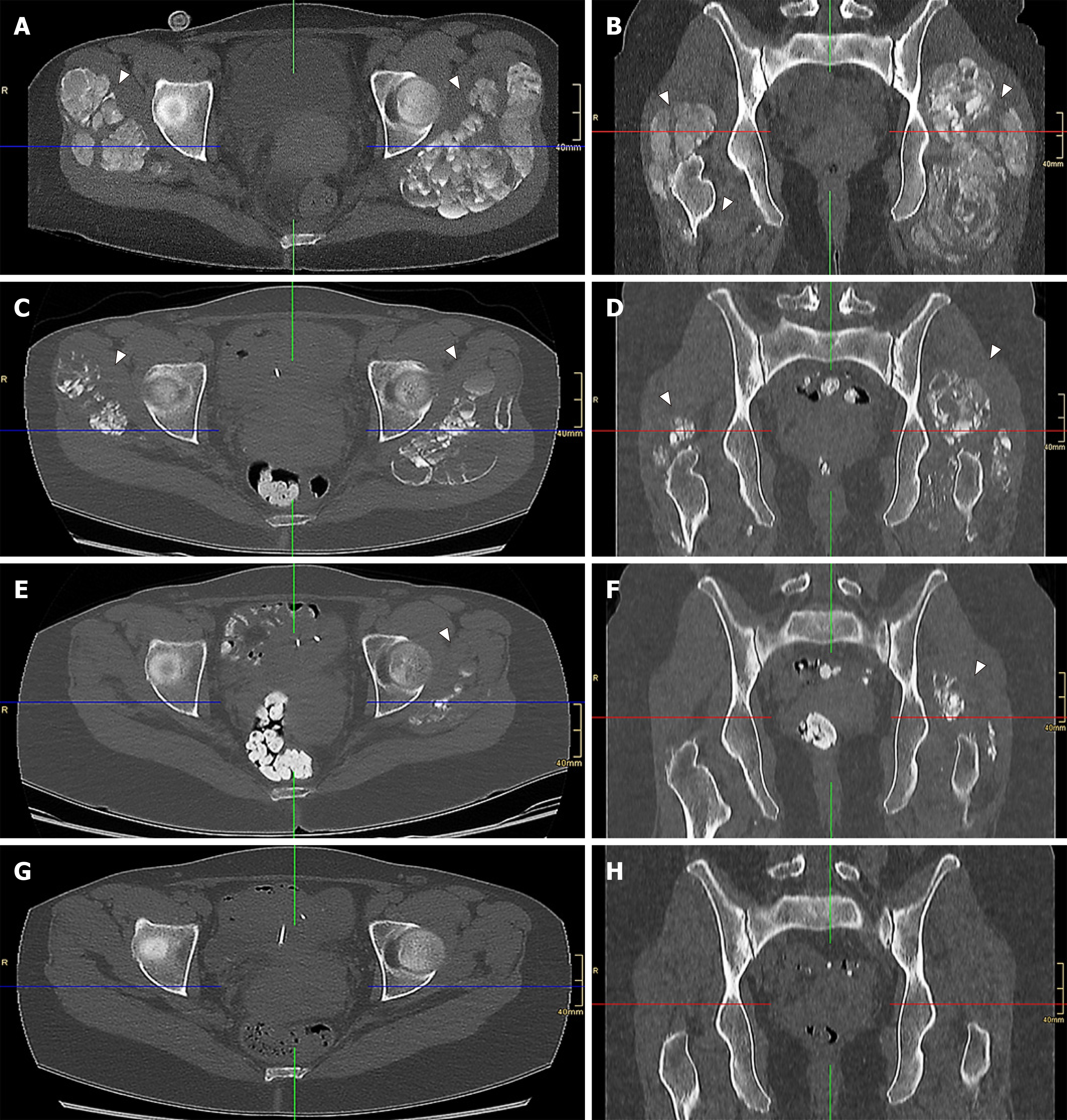
Figure 1 Computed tomography images of severe tumoral calcinosis with complete remission in an end-stage renal disease patient.
A, B: Initial diagnosis; C, D: At 2 mo; E, F: At 4 mo; G, H: At 11 mo. Computed tomography scan shows severe tumoral calcinosis of the trochanter major region depicting extensive periarticular cyst-like calcified space-consuming lesions (arrow heads) at initial diagnosis (A, B) and their continuous remission thereafter at 2 mo (C, D), 4 mo (E, F) and 11 mo (G, H) after the initial diagnosis at which point complete remission was achieved (the blue lines represent the respective frontal plane in the transversal plane images, the red lines represent the respective transversal plane in the frontal plane images, and the green lines represent the median sagittal plane in all images).
- Citation: Westermann L, Isbell LK, Breitenfeldt MK, Arnold F, Röthele E, Schneider J, Widmeier E. Recuperation of severe tumoral calcinosis in a dialysis patient: A case report. World J Clin Cases 2019; 7(23): 4004-4010
- URL: https://www.wjgnet.com/2307-8960/full/v7/i23/4004.htm
- DOI: https://dx.doi.org/10.12998/wjcc.v7.i23.4004


