Copyright
©The Author(s) 2019.
World J Clin Cases. Nov 26, 2019; 7(22): 3895-3903
Published online Nov 26, 2019. doi: 10.12998/wjcc.v7.i22.3895
Published online Nov 26, 2019. doi: 10.12998/wjcc.v7.i22.3895
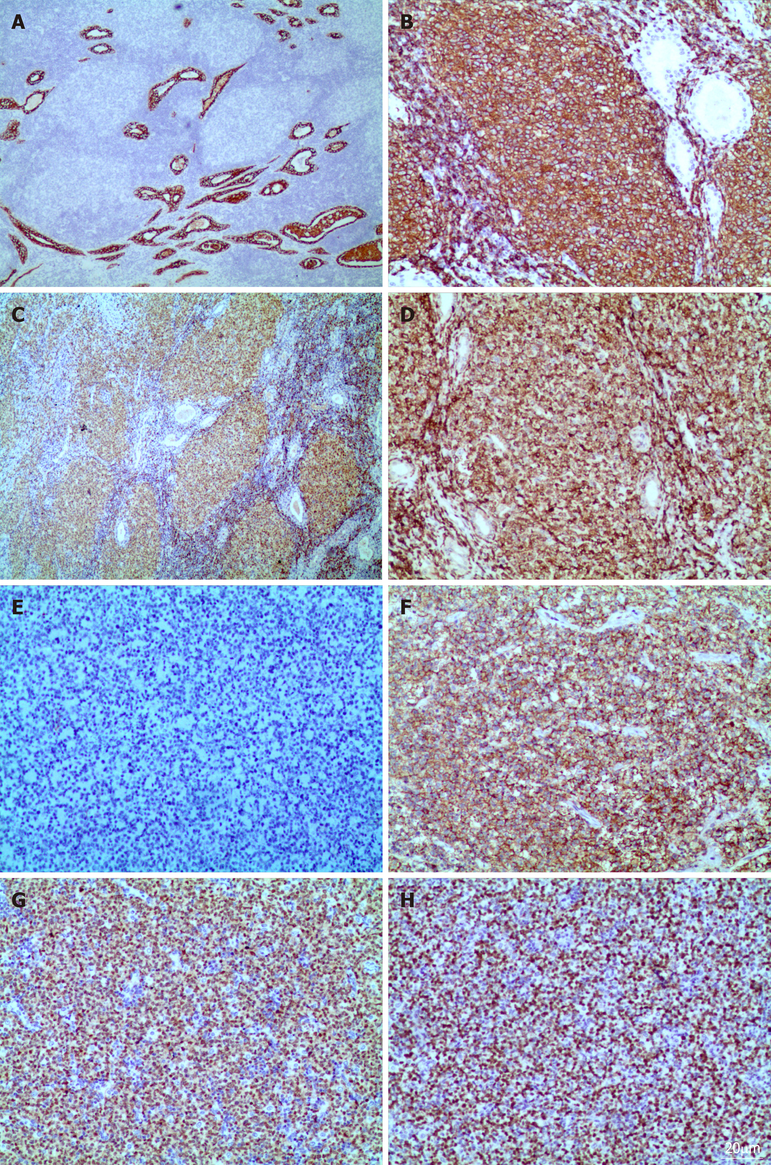
Figure 2 Immunotype of lymphoma with Warthin’s tumor.
A: The columnar oncocytic cells were positive for AE1/AE3 (Magnification ×100). B-E: The neoplastic lymphoid cells which were located incoarctate follicular were positive for CD20 (B), Pax-5 (C), bcl-2 (D), negative for CD10 (E); F, G: The adjacent diffusely medium- to large- size lymphoid cells was positive for CD20 (F), MUM-1(G); H: The Ki-67 proliferation index was estimated at approximately 80%. (Immunohistochemical stain, Magnification ×200).
- Citation: Wang CS, Chu X, Yang D, Ren L, Meng NL, Lv XX, Yun T, Cao YS. Diffuse large B-cell lymphoma arising from follicular lymphoma with warthin’s tumor of the parotid gland - immunophenotypic and genetic features: A case report. World J Clin Cases 2019; 7(22): 3895-3903
- URL: https://www.wjgnet.com/2307-8960/full/v7/i22/3895.htm
- DOI: https://dx.doi.org/10.12998/wjcc.v7.i22.3895


