Copyright
©The Author(s) 2019.
World J Clin Cases. Nov 26, 2019; 7(22): 3844-3850
Published online Nov 26, 2019. doi: 10.12998/wjcc.v7.i22.3844
Published online Nov 26, 2019. doi: 10.12998/wjcc.v7.i22.3844
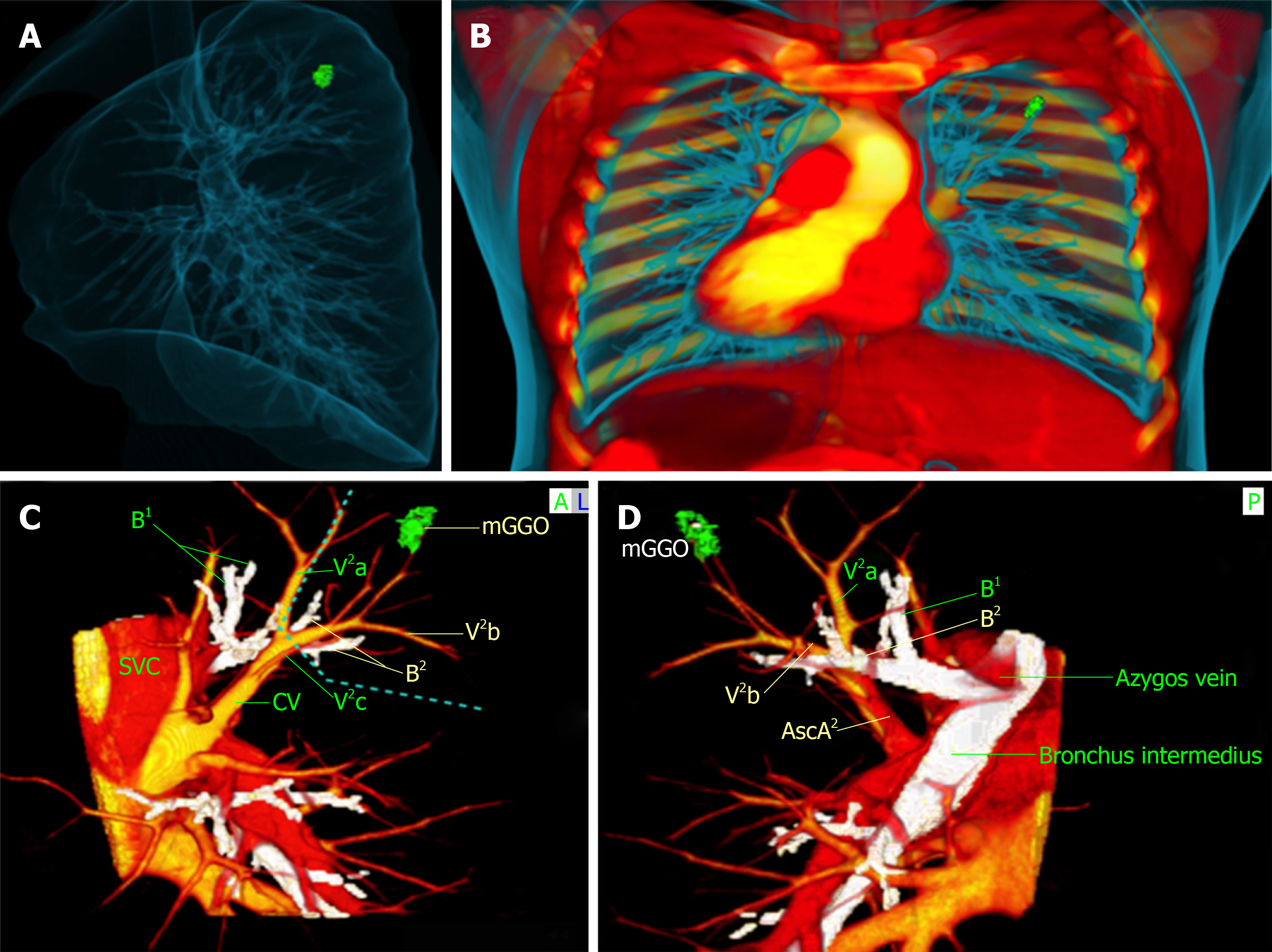
Figure 3 Three-dimensional images reconstructed with OsiriX software.
A and B: Three-dimensional structure and spatial relation of bronchi and blood vessels; C and D: Exact three-dimensional computed tomography bronchography and angiography relationships between the mixed ground-glass opacity (mGGO) lesion and pulmonary anatomical structures. Green: The mGGO lesion; Yellow: Pulmonary veins; Red: Pulmonary artery; White: Bronchus. The blue dotted line denotes the safety margin. 3D-CTBA: Three-dimensional computed tomography bronchography and angiography; mGGO: Mixed ground-glass opacity.
- Citation: Wu YJ, Bao Y, Wang YL. Thoracoscopic segmentectomy assisted by three-dimensional computed tomography bronchography and angiography for lung cancer in a patient living with situs inversus totalis: A case report. World J Clin Cases 2019; 7(22): 3844-3850
- URL: https://www.wjgnet.com/2307-8960/full/v7/i22/3844.htm
- DOI: https://dx.doi.org/10.12998/wjcc.v7.i22.3844


