Copyright
©The Author(s) 2019.
World J Clin Cases. Nov 26, 2019; 7(22): 3832-3837
Published online Nov 26, 2019. doi: 10.12998/wjcc.v7.i22.3832
Published online Nov 26, 2019. doi: 10.12998/wjcc.v7.i22.3832
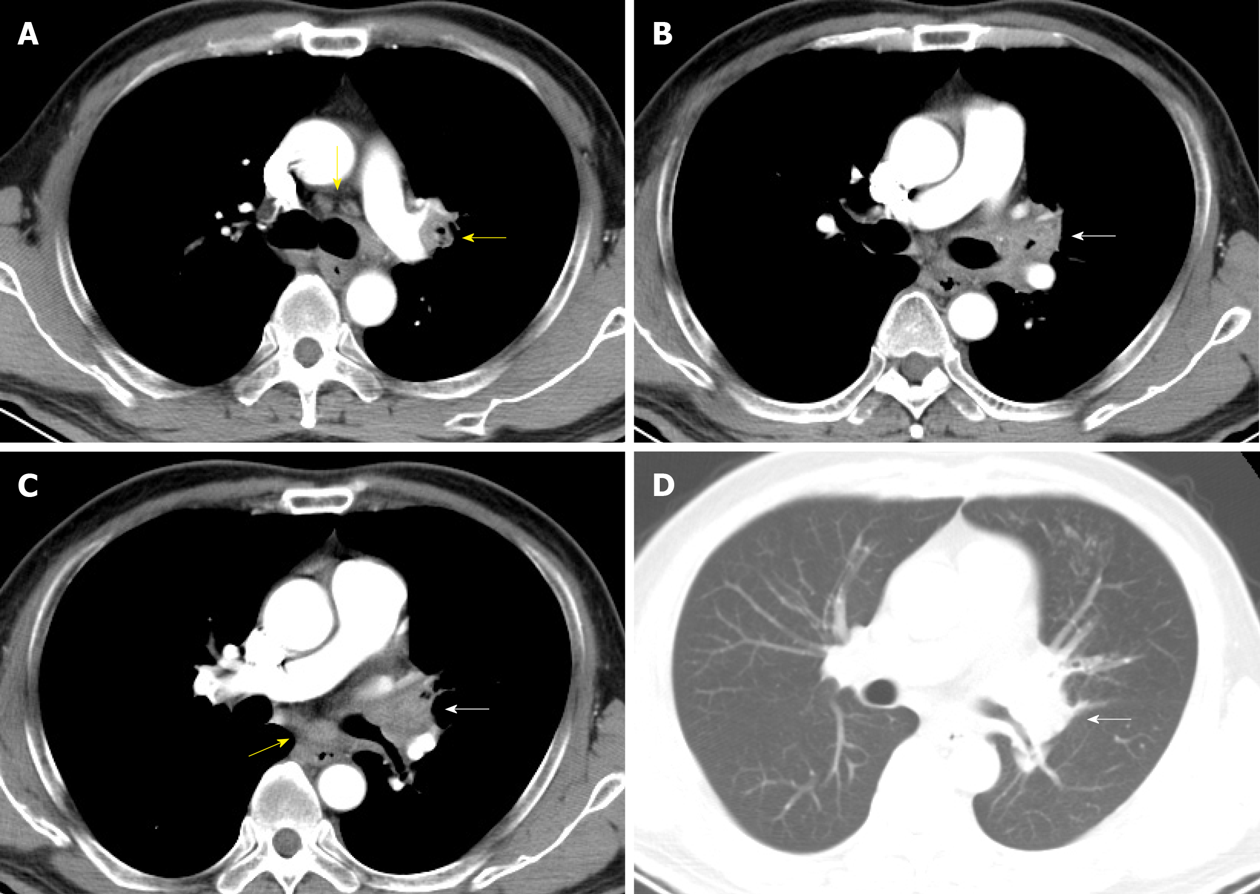
Figure 1 Contrast-enhanced chest computed tomography images (2018-04-07).
A-C: Mediastinal window; D: Lung window. Contrast-enhanced chest computed tomography scan revealed enlarged mediastinum and hilum lymph nodes (yellow arrow) and an enlarged left hilum with a mass-like lesion leading to stenosis of the proximal part of the left upper bronchus (white arrow).
- Citation: Su SS, Zhou Y, Xu HY, Zhou LP, Chen CS, Li YP. Invasive aspergillosis presenting as hilar masses with stenosis of bronchus: A case report. World J Clin Cases 2019; 7(22): 3832-3837
- URL: https://www.wjgnet.com/2307-8960/full/v7/i22/3832.htm
- DOI: https://dx.doi.org/10.12998/wjcc.v7.i22.3832


