
















©The Author(s) 2019.
World J Clin Cases. Nov 6, 2019; 7(21): 3639-3648
Published online Nov 6, 2019. doi: 10.12998/wjcc.v7.i21.3639
Published online Nov 6, 2019. doi: 10.12998/wjcc.v7.i21.3639
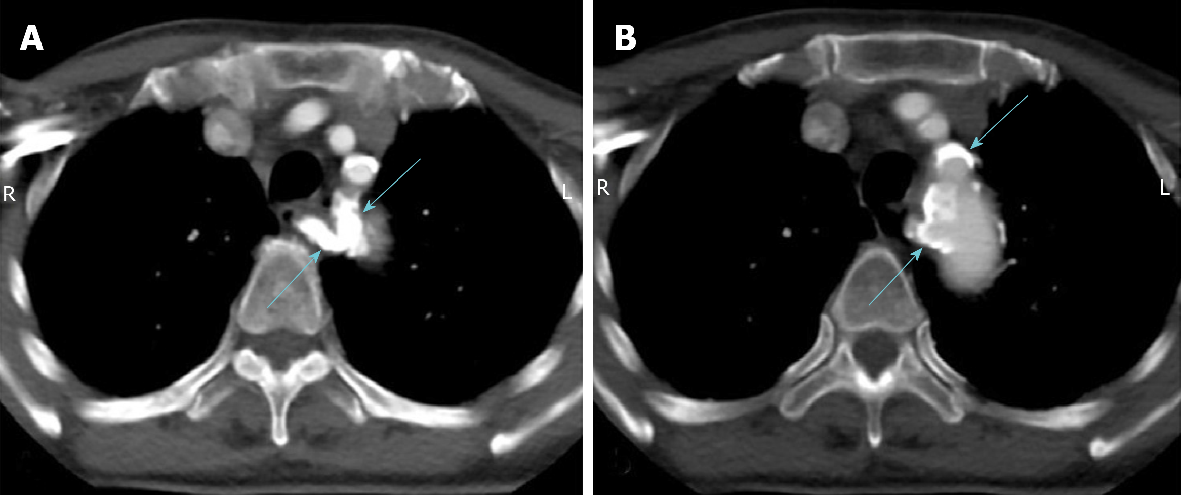
Figure 7 Enhanced computed tomography.
A, B: Most of the artery wall calcification plaques were concentrated at the beginning of the aortic branch artery, especially the aberrant right subclavian artery (blue arrow), which led to the aberrant right subclavian steal syndrome.
- Citation: Sun YY, Zhang GM, Zhang YB, Du X, Su ML. Bilateral common carotid artery common trunk with aberrant right subclavian artery combined with right subclavian steal syndrome: A case report. World J Clin Cases 2019; 7(21): 3639-3648
- URL: https://www.wjgnet.com/2307-8960/full/v7/i21/3639.htm
- DOI: https://dx.doi.org/10.12998/wjcc.v7.i21.3639