Copyright
©The Author(s) 2019.
World J Clin Cases. Oct 26, 2019; 7(20): 3322-3328
Published online Oct 26, 2019. doi: 10.12998/wjcc.v7.i20.3322
Published online Oct 26, 2019. doi: 10.12998/wjcc.v7.i20.3322
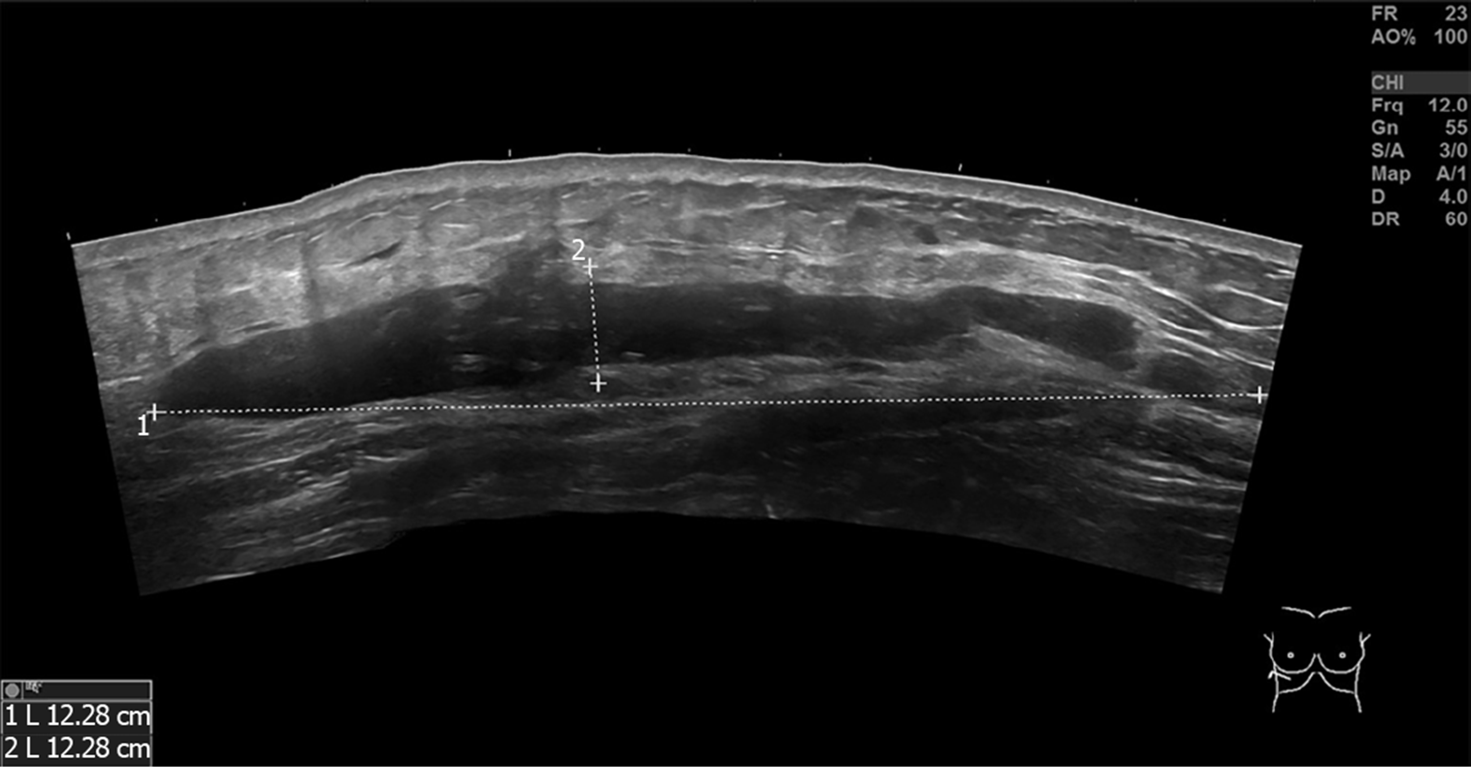
Figure 6 Ultrasonography of the bilateral hypochondriac region demonstrating that there was a hypoechoic area shaped like a bar on each side.
- Citation: Zhang MX, Li SY, Xu LL, Zhao BW, Cai XY, Wang GL. Repeated lumps and infections: A case report on breast augmentation complications. World J Clin Cases 2019; 7(20): 3322-3328
- URL: https://www.wjgnet.com/2307-8960/full/v7/i20/3322.htm
- DOI: https://dx.doi.org/10.12998/wjcc.v7.i20.3322


