Copyright
©The Author(s) 2019.
World J Clin Cases. Oct 26, 2019; 7(20): 3322-3328
Published online Oct 26, 2019. doi: 10.12998/wjcc.v7.i20.3322
Published online Oct 26, 2019. doi: 10.12998/wjcc.v7.i20.3322
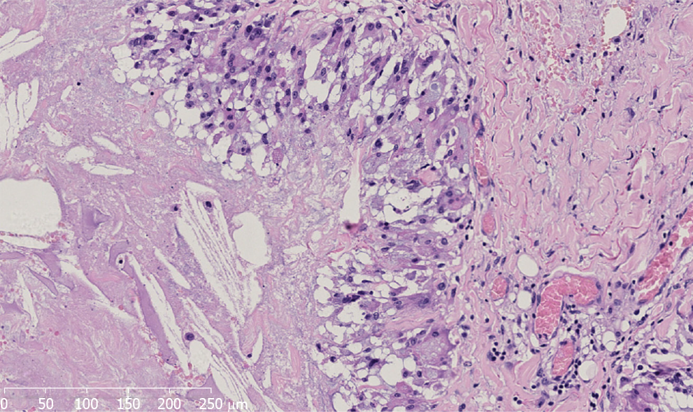
Figure 4 Microscopic appearance of the gray-white and gray-yellow tissue revealed dilated or fissured interconnected cysts with pseudopapillary structures.
The cyst wall was composed of fibrous tissue with hyaline degeneration. There are many tissue cells and foreign body giant cells in the inner wall (hematoxylin and eosin staining; original magnification: 200×).
- Citation: Zhang MX, Li SY, Xu LL, Zhao BW, Cai XY, Wang GL. Repeated lumps and infections: A case report on breast augmentation complications. World J Clin Cases 2019; 7(20): 3322-3328
- URL: https://www.wjgnet.com/2307-8960/full/v7/i20/3322.htm
- DOI: https://dx.doi.org/10.12998/wjcc.v7.i20.3322


