Published online Oct 26, 2019. doi: 10.12998/wjcc.v7.i20.3372
Peer-review started: May 27, 2019
First decision: August 1, 2019
Revised: August 22, 2019
Accepted: September 13, 2019
Article in press: September 12, 2019
Published online: October 26, 2019
Ewing’s sarcoma (ES) is regarded as a skeletal tumor, with few instances of extra-skeletal ES. A primary ES in the ureter is extremely rare.
We report the case of a 69-year-old woman who presented with intermittent flank pain and hematuria and was found to have a mass in the left ureter. Pathology of the excised mass indicated ES. The clinical treatment and pathologic characteristics in this case, and a review of the literature describing ES in the urinary system, are presented.
Due to the rarity and malignancy of ES in ureter, early diagnosis and prompt surgical treatment are critical.
Core tip: Extra-skeletal Ewing’s sarcomas (EES) are rare aggressive malignant small round cell tumors. To date, there have been only two reported case of Ewing’s sarcoma of the ureter including the patient presented in this report. The patient presented in this report is a 69-year-old woman with intermittent flank pain and hematuria.
- Citation: Li XX, Bi JB. Ureteral Ewing’s sarcoma in an elderly woman: A case report. World J Clin Cases 2019; 7(20): 3372-3376
- URL: https://www.wjgnet.com/2307-8960/full/v7/i20/3372.htm
- DOI: https://dx.doi.org/10.12998/wjcc.v7.i20.3372
Ewing’s sarcoma (ES) is a rare malignant disease in which tumor cells are usually found in the bone or soft tissue and typically occurs between the ages of 10 years and 20 years[1]. ES arises primarily from bone and is rarely of extra-skeletal origin[2]. Of the extraosseous ES reported, to our knowledge ureteral (U)ES has only been noted in a 17-year-old girl[3]. We present the clinical and pathologic features of an elderly woman who was diagnosed with UES and discuss the differences between this patient and the 17-year-old patient with UES.
A 69-year-old woman presented with a 10-d history of intermittent left flank pain.
Following impact, she felt frequent urination (five to six times in the day and three times in the night). Past medical history showed hematuria 1 year ago.
There were no significant comorbidities at admission.
The patient was a non-smoker, without personal or family history of other diseases.
No obvious abnormalities in the physical examination.
The results of laboratory studies showed poor renal function with increased creatinine (271 μmol/L) and urea (12.98 mmol/L).
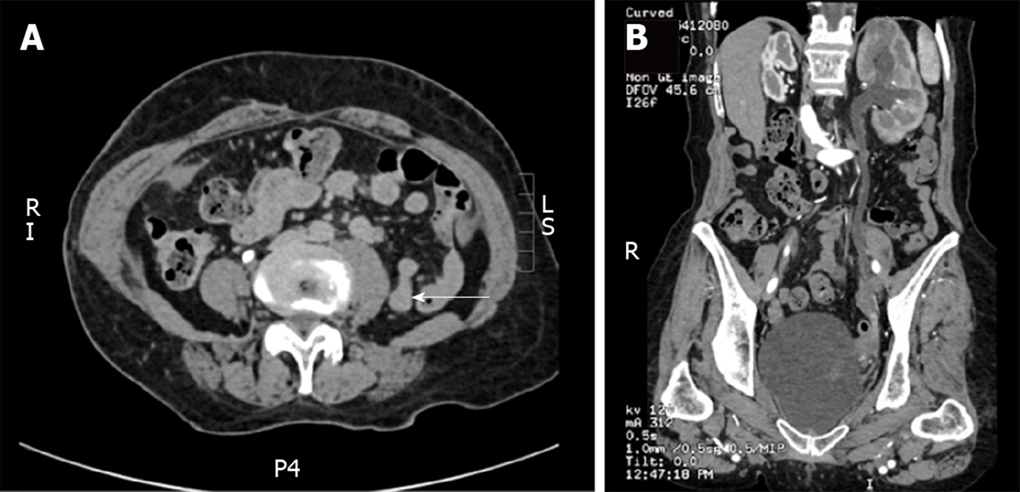
Computed tomography (Figure 1) revealed a 1.5 cm soft tissue mass in the left ureteral pelvic segment with left hydronephrosis and ureter dilatation.
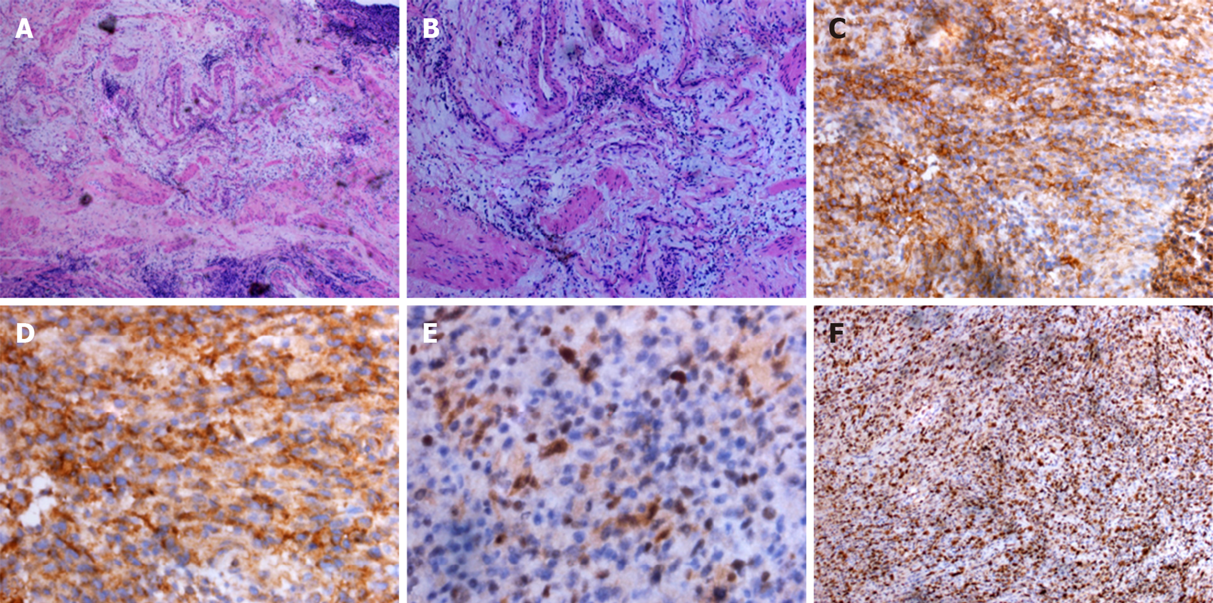
Histopathology examination (Figure 2) revealed a pT2N0M0 tumor 1.5 cm in size composed of small cells with indistinct cell borders, scant cytoplasm and atypia nuclei. Immunohistochemistry demonstrated that the tumor cells were positive for cluster of differentiation 99 (CD99), transducin-like enhancer protein 1, and 70% were positive for Ki67. The pathology department of our hospital diagnosed it to be ES, with no metastasis of para-vascular lymph nodes.
The patient underwent resection of the ureter lesion, end anastomosis and local lymph node dissection.
When taking into account the patient’s age and cardiopulmonary function, she was not treated with chemotherapy. Four weeks after surgery, her creatinine level was normal. Three months later, the patient underwent re-examination with computed tomography scans of the chest, abdomen and pelvis, and no recurrence or metastases were found. The patient is still alive.
ES is a rare entity. Similar to other sarcomas, ES is a monomorphic small round cell tumor that originates from the neural crest and accounts for 10% of all sarcomas[4]. Individual cases of ES have been reported in almost all organs in the urinary system.
Primary ES in the kidney was first reported by Mor et al[5] in 1994, and this was the earliest discovery of ES in the urinary system. Over the past 15 years, the incidence of renal ES has steadily increased with over 100 cases reported in the literature[6], and Risi et al[7] noted 116 cases of renal ES. The majority of patients were men (55%), and the median age was 28.0 years (20.0-42.0 years). All patients had clinical symptoms as the initial presentation of disease. Pain was the most frequent symptom (54%), followed by hematuria (29%). The median overall survival (OS) was 26.5 mo. Thirty-three percent of these patients were found to have metastasis when the disease was diagnosed. The median OS of metastatic patients was 24 mo, and the three most frequent sites of metastases were lung (60%), liver (37%) and abdominal wall lymph nodes (20%). Patients with metastatic disease have more than a 4-fold increase in the relative risk of death compared to non-metastatic patients at diagnosis.
Of all the cases reported, 89% of patients underwent surgery. The 2-year survival rate was 80% compared with 30% in those who did not undergo surgery. Half of patients also underwent preoperative or postoperative chemotherapy. The 12-mo OS was 93% compared to 75% in those who did not undergo chemotherapy.
The diagnosis of ES mainly depends on histopathology and immunohistochemistry of surgical specimens. ES cells are typically positive for CD99 and friend leukemia integration 1 transcription factor (FLI-1) in more than 90% of cases. CD99 has been found to be nonspecific in the diagnosis of ES; thus, the presence of other typical biomarkers including SYN (synaptophysin), FLI-1 and Ki67 should be determined[8,9]. Clinically, renal ES patients treated with seven cycles of chemotherapy including vincristine, cyclophosphamide, dactinomycin and doxorubicin after radical nephrectomy remained disease-free for 7.5 years. Moreover, postoperative metastases to the liver and lymph nodes were treated with chemotherapy (six cycles of vincristine, doxorubicin and cyclophosphamide), and the response rate was approximately 90% based on the RECIST guidelines[10].
Lam et al[11] described a 31-year-old woman diagnosed with bladder ES who underwent complete resection of the bladder after 7 cycles of neoadjuvant chemotherapy, which reduced the size of the tumor from 8.1 cm to 2.5 cm, confirming the effectiveness of this therapeutic strategy. These authors also gathered information on all patients with bladder ES in the English literature. In these 13 cases with bladder ES, patient age ranged from 10 years to 81 years, with a mean age of 41 years. The most common presenting symptom was hematuria, followed by dysuria. Immunohistochemical findings showed that MIC2 was present in 100% of patients, whereas desmin was present in only 5 patients. This study also found that approximately 50% of patients had EWS/FLI-1 fusion following molecular genetics analysis. At the time of diagnosis, 2 patients were identified as having metastases, including pulmonary and abdominal wall metastases. The treatment of bladder ES was based on the chemotherapy regimen used for ES[12].
According to the literature review, only 10 cases of prostate ES have been reported since 2003. The survival rate of these patients was 2 to 12 mo. The treatment strategy included surgery and adjuvant and neoadjuvant chemotherapy, but there was limited data acquisition[13].
ES occurring in the ureter was first reported in 2003 by Charny et al[3] in a 17-year-old girl who presented with flank pain and hematuria. During surgery, a mass was excised from the wall of the right ureter followed by anastomosis of the ureter. The t (11; 22) chromosomal translocation specific to ES was confirmed by reverse transcriptase polymerase chain reaction. Our patient was also diagnosed with UES. When comparing the two patients, our patient was older and did not fit into the typical age group for the occurrence of ES. Renal function in this patient was seriously impaired, and it was necessary to remove the obstruction, otherwise the patient would have developed renal failure. The previously reported case was discharged from hospital 4 days after surgery, and long-term follow-up information could not be obtained. Thus, the patient's surgical outcome could not be assessed. However, in our patient, postoperative renal function returned to normal, and no recurrence or metastases were found in the subsequent 6 mo. These findings confirmed that surgical resection was effective for the initial treatment of UES. The previously reported patient with UES was young, and her cardiopulmonary function allowed her to tolerate chemotherapy. However, as our patient was elderly, her physical condition prevented her from receiving other forms of treatment after surgery. Adjuvant chemotherapy may be beneficial in patients with ES occurring at other sites in the urinary system and may improve their survival time.
We describe an extremely rare UES in an elderly woman treated with resection of the lesion and end anastomosis. Pathological analysis and immunohistochemistry of the surgical specimen were performed to confirm the diagnosis. Due to the rarity and malignancy of this tumor, early diagnosis and prompt surgical treatment are critical.
Manuscript source: Unsolicited manuscript
Specialty type: Medicine, Research and Experimental
Country of origin: China
Peer-review report classification
Grade A (Excellent): A
Grade B (Very good): 0
Grade C (Good): C
Grade D (Fair): 0
Grade E (Poor): 0
P-Reviewer: Chetty R, Sureshkumar K S-Editor: Dou Y L-Editor: Filipodia E-Editor: Liu JH
| 1. | Llombart-Bosch A, Machado I, Navarro S, Bertoni F, Bacchini P, Alberghini M, Karzeladze A, Savelov N, Petrov S, Alvarado-Cabrero I, Mihaila D, Terrier P, Lopez-Guerrero JA, Picci P. Histological heterogeneity of Ewing's sarcoma/PNET: an immunohistochemical analysis of 415 genetically confirmed cases with clinical support. Virchows Arch. 2009;455:397-411. [PubMed] [DOI] [Cited in This Article: ] [Cited by in Crossref: 154] [Cited by in F6Publishing: 138] [Article Influence: 9.2] [Reference Citation Analysis (0)] |
| 2. | Ding Y, Huang Z, Ding Y, Jia Z, Gu C, Xue R, Yang J. Primary Ewing's Sarcoma/Primitive Neuroectodermal Tumor of Kidney with Caval Involvement in a Pregnant Woman. Urol Int. 2016;97:365-368. [PubMed] [DOI] [Cited in This Article: ] [Cited by in Crossref: 2] [Cited by in F6Publishing: 3] [Article Influence: 0.4] [Reference Citation Analysis (0)] |
| 3. | Charny CK, Glick RD, Genega EM, Meyers PA, Reuter VE, La Quaglia MP. Ewing's sarcoma/primitive neuroectodermal tumor of the ureter: a case report and review of the literature. J Pediatr Surg. 2000;35:1356-1358. [PubMed] [DOI] [Cited in This Article: ] [Cited by in Crossref: 18] [Cited by in F6Publishing: 20] [Article Influence: 0.8] [Reference Citation Analysis (0)] |
| 4. | Funahashi Y, Hattori R, Yamamoto T, Mizutani K, Yoshino Y, Matsukawa Y, Sassa N, Okumura K, Gotoh M. Ewing's sarcoma / primitive neuroectodermal tumor of the kidney. Aktuelle Urol. 2009;40:247-249. [PubMed] [DOI] [Cited in This Article: ] [Cited by in Crossref: 8] [Cited by in F6Publishing: 9] [Article Influence: 0.6] [Reference Citation Analysis (0)] |
| 5. | Mor Y, Nass D, Raviv G, Neumann Y, Nativ O, Goldwasser B. Malignant peripheral primitive neuroectodermal tumor (PNET) of the kidney. Med Pediatr Oncol. 1994;23:437-440. [PubMed] [DOI] [Cited in This Article: ] [Cited by in Crossref: 43] [Cited by in F6Publishing: 52] [Article Influence: 1.7] [Reference Citation Analysis (0)] |
| 6. | Rowe RG, Thomas DG, Schuetze SM, Hafez KS, Lawlor ER, Chugh R. Ewing sarcoma of the kidney: case series and literature review of an often overlooked entity in the diagnosis of primary renal tumors. Urology. 2013;81:347-353. [PubMed] [DOI] [Cited in This Article: ] [Cited by in Crossref: 24] [Cited by in F6Publishing: 25] [Article Influence: 2.3] [Reference Citation Analysis (0)] |
| 7. | Risi E, Iacovelli R, Altavilla A, Alesini D, Palazzo A, Mosillo C, Trenta P, Cortesi E. Clinical and pathological features of primary neuroectodermal tumor/Ewing sarcoma of the kidney. Urology. 2013;82:382-386. [PubMed] [DOI] [Cited in This Article: ] [Cited by in Crossref: 44] [Cited by in F6Publishing: 48] [Article Influence: 4.4] [Reference Citation Analysis (0)] |
| 8. | Celli R, Cai G. Ewing Sarcoma/Primitive Neuroectodermal Tumor of the Kidney: A Rare and Lethal Entity. Arch Pathol Lab Med. 2016;140:281-285. [PubMed] [DOI] [Cited in This Article: ] [Cited by in Crossref: 17] [Cited by in F6Publishing: 19] [Article Influence: 2.4] [Reference Citation Analysis (0)] |
| 9. | Antonescu C. Round cell sarcomas beyond Ewing: emerging entities. Histopathology. 2014;64:26-37. [PubMed] [DOI] [Cited in This Article: ] [Cited by in Crossref: 132] [Cited by in F6Publishing: 133] [Article Influence: 12.1] [Reference Citation Analysis (0)] |
| 10. | Castro EC, Parwani AV. Ewing sarcoma/primitive neuroectodermal tumor of the kidney: two unusual presentations of a rare tumor. Case Rep Med. 2012;2012:190581. [PubMed] [DOI] [Cited in This Article: ] [Cited by in Crossref: 8] [Cited by in F6Publishing: 11] [Article Influence: 0.9] [Reference Citation Analysis (0)] |
| 11. | Lam CJ, Shayegan B. Complete resection of a primitive neuroectodermal tumour arising in the bladder of a 31-year-old female after neoadjuvant chemotherapy. Can Urol Assoc J. 2016;10:E264-E267. [PubMed] [DOI] [Cited in This Article: ] [Cited by in Crossref: 5] [Cited by in F6Publishing: 6] [Article Influence: 0.8] [Reference Citation Analysis (0)] |
| 12. | Grier HE, Krailo MD, Tarbell NJ, Link MP, Fryer CJ, Pritchard DJ, Gebhardt MC, Dickman PS, Perlman EJ, Meyers PA, Donaldson SS, Moore S, Rausen AR, Vietti TJ, Miser JS. Addition of ifosfamide and etoposide to standard chemotherapy for Ewing's sarcoma and primitive neuroectodermal tumor of bone. N Engl J Med. 2003;348:694-701. [PubMed] [DOI] [Cited in This Article: ] [Cited by in Crossref: 942] [Cited by in F6Publishing: 860] [Article Influence: 41.0] [Reference Citation Analysis (0)] |
| 13. | Esch L, Barski D, Bug R, Otto T. Prostatic sarcoma of the Ewing family in a 33-year-old male - A case report and review of the literature. Asian J Urol. 2016;3:103-106. [PubMed] [DOI] [Cited in This Article: ] [Cited by in Crossref: 5] [Cited by in F6Publishing: 5] [Article Influence: 0.6] [Reference Citation Analysis (0)] |


