
















©The Author(s) 2019.
World J Clin Cases. Jan 26, 2019; 7(2): 228-235
Published online Jan 26, 2019. doi: 10.12998/wjcc.v7.i2.228
Published online Jan 26, 2019. doi: 10.12998/wjcc.v7.i2.228
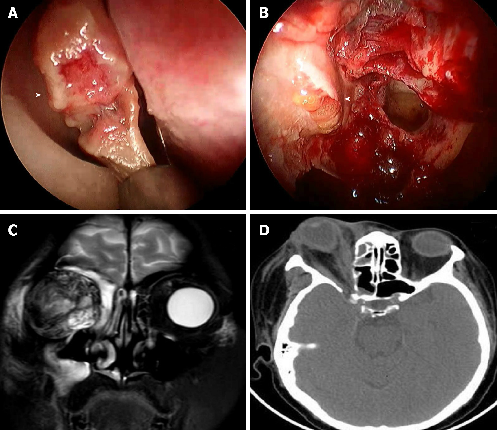
Figure 1 Nasal endoscopy, computed tomography, and magnetic resonance imaging images.
A: Endoscopic sinus surgery showed that the middle turbinate turned to be necrotic and fragile; B: The orbital fat appeared dark yellow, losing its bright color; C: Magnetic resonance imaging T2-weighted coronal image; D: Axial computed tomography scan with soft tissue window revealed inflammatory changes in the orbital fat, right ocular proptosis, thickening of extraocular muscles, and the distorted eyeball.
- Citation: Liu YC, Zhou ML, Cheng KJ, Zhou SH, Wen X, Chang CD. Successful treatment of invasive fungal rhinosinusitis caused by Cunninghamella: A case report and review of the literature. World J Clin Cases 2019; 7(2): 228-235
- URL: https://www.wjgnet.com/2307-8960/full/v7/i2/228.htm
- DOI: https://dx.doi.org/10.12998/wjcc.v7.i2.228